Session: MP31: Renal Transplantation & Vascular Surgery I
MP31-02: In the Era of Computed Tomography Volumetry: is Isotope Renography Still Routinely Required in the Preparation and Selection of Living Kidney Donors?
Professor of Urology Urology and Nephrology Center, Faculty of Medicine, Mansoura University
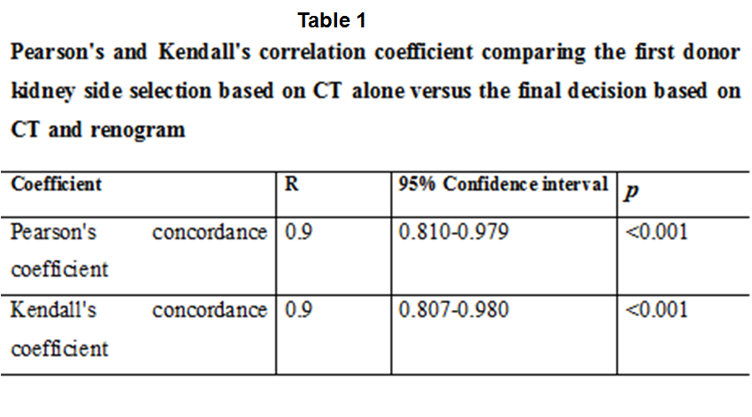
Introduction: Side selection in living kidney donors (LKD) before renal transplantation (RT) depends mainly on the split-renal function (SRF) assessed by isotope renography and structural anatomy of the kidney determined by computed tomography angiography (CTA). If we can use a single modality to assess both, we can reduce time and cost. The objective is to compare the CT-split renal volume (CT-SRV) to the SRF derived from DTPA Renography with regard to the decision of donor side selection. Methods: The study was performed between July 2017 and July 2020. In potential donors, renal volume was measured using computer generated volumetric scanning system. CT-SRV was estimated by dividing one kidney volume by the volume of both. Similarly, SRF was estimated by glomerular filtration rate (GFR) on renogram. Donor side selection was made by multi-disciplinary team. After complete evaluation, donor side selection was determined by CT alone (SRV and vascular anatomy). Then, renogram was shown and selection made by CT alone was compared to that based on CT and renogram. Results: 258 consecutive donors (94 males and 164 females; mean age 42±9.7 years) were included. SRF and SRV were comparable in 177 (Rt) & 172 (Lt) cases. Using CT alone, the right and left kidney were chosen in 91 and 167. Using CT and renogram, 83 and 175 right and left kidneys were selected with mean GFR of 55.2±8.9 ml/min and CT volume 156±28 mL. The choice of the donor side based on CT alone was in agreement with the choice based on both CT and renogram in 237 (92%), with a difference in choice in 21 cases (8%) (table 1). Subset analysis of these 21 cases showed that 15 had multiple vessels, small cysts and/ or gravels and all had a significant disparity (>5.3%) on CT volumetry. Conclusions: Regarding donor side selection, CT volumetry has high concordance (>90%) with isotope renography. It is, therefore, considered a sufficient pre-transplant single diagnostic tool for this purpose. However, if significant disparity in SRV is diagnosed by CT or in the cases with multiple vessels, cysts and/ or gravels, complete evaluation using both CT and isotope renography should be conducted SOURCE OF Funding: None