Back
Abstract Session
Session: Abstracts: Imaging of Rheumatic Diseases (2204–2208)
2208: Diagnostic Performance of Hip Ultrasound in Calcium Pyrophosphate Deposition Disease
Monday, November 14, 2022
4:00 PM – 4:10 PM Eastern Time
Location: Room 126
.png)
Carlos Pineda, PhD
Instituto Nacional de Rehabilitacion
Mexico City, Federal District, Mexico
Presenting Author(s)
Carina Soto-Fajardo1, Fabian Carranza-Enriquez2, Sinthia Solorzano Flores2, Abish Angeles2, Paola Flores2, Raul Pichardo-Bahena2, Luis Javier Jara Quezada2, Angélica Peña Ayala2, Victor-Manuel Ilizaliturri-Sanchez2, Georgios Filippou3 and Carlos Pineda2, 1Instituto Nacional de Rehabilitacion \"Luis Guillermo Ibarra Ibarra\", Ciudad de México, Mexico, 2Instituto Nacional de Rehabilitacion, Ciudad de México, Mexico, 3Rheumatology Department, Luigi Sacco University Hospital, Siena, Italy
Background/Purpose: Calcium Pyrophosphate Deposition Disease (CPPD) is a chronic and potentially incapacitating disease. The reference standard for its diagnosis is the identification of calcium pyrophosphate (CPP) crystals in synovial fluid. Ultrasound (US) has been proven to be a high sensibility and specificity tool for the diagnosis of CPPD, but it has not yet been determined its diagnostic performance for hip involvement. The aim of this research is to evaluate the diagnostic performance of US compared with synovial fluid analysis and histopathology (hyaline cartilage, fibrocartilage, synovial membrane) for the identification of hip CPP deposits.
Methods: Patients older than 50 years old with osteoarthritis programed for hip replacement surgery in a referral hospital. Patients with a primary rheumatologic disease were excluded. A pre-surgical US of the affected hip was performed with a LOGIQTMe device and a convex transducer (2-5 MHz); the anatomical structures evaluated were the acetabular fibrocartilage (FC) and the hyaline cartilage of the femoral head (HC), video tracking was recorded and a dichotomic score was assigned to determine the presence or absence of CPP deposits (in line with OMERACT definitions). During surgery, a sample of hip synovial fluid was obtained and examined using polarized light microscopy. After the surgery an experimented pathologist examined the FC and HC in search for CPP crystals.
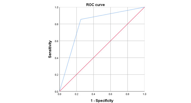
Results: 67 patients were included, 55.2% were women with a mean age of 64±7.9 years. A frequency of 82% (55/67) of CPP deposits was found through US examination, of which 29/67 (43.3%) where found on FC and 42/67(62.7%) were found on HC. Regarding pathology evaluation, a prevalence of 93.9% of CPP crystals was found, 83.6% (56/67) were found on FC and 86.6% (58/67) on HC. 24% of patients had synovial effusion and 3% had synovial hypertrophy. Synovial fluid was obtained in 64.2% (43/67) of patients, with a median volume of 0.8mL (IQR 0.45-1.15mL), CPP crystals were found in 14.9% (10/43) of samples. Chondrocalcinosis in XR was found in 14.9% (10/67). A sensitivity of 85% and a specificity of 75% were obtained, the positive predictive value was 98% and the negative predictive value was 25%. The area under the curve for US compared with histopathology for the diagnosis of hip CPPD was 0.80 (CI 95% 0.55-1) (Figure 1).
Conclusion: US is a valid imaging method with good diagnostic performance for the diagnosis of CPPD in the coxofemoral joint.
 Figure 1. ROC curve of US
Disclosures: C. Soto-Fajardo, None; F. Carranza-Enriquez, None; S. Solorzano Flores, None; A. Angeles, None; P. Flores, None; R. Pichardo-Bahena, None; L. Jara Quezada, None; A. Peña Ayala, None; V. Ilizaliturri-Sanchez, None; G. Filippou, None; C. Pineda, None.
Background/Purpose: Calcium Pyrophosphate Deposition Disease (CPPD) is a chronic and potentially incapacitating disease. The reference standard for its diagnosis is the identification of calcium pyrophosphate (CPP) crystals in synovial fluid. Ultrasound (US) has been proven to be a high sensibility and specificity tool for the diagnosis of CPPD, but it has not yet been determined its diagnostic performance for hip involvement. The aim of this research is to evaluate the diagnostic performance of US compared with synovial fluid analysis and histopathology (hyaline cartilage, fibrocartilage, synovial membrane) for the identification of hip CPP deposits.
Methods: Patients older than 50 years old with osteoarthritis programed for hip replacement surgery in a referral hospital. Patients with a primary rheumatologic disease were excluded. A pre-surgical US of the affected hip was performed with a LOGIQTMe device and a convex transducer (2-5 MHz); the anatomical structures evaluated were the acetabular fibrocartilage (FC) and the hyaline cartilage of the femoral head (HC), video tracking was recorded and a dichotomic score was assigned to determine the presence or absence of CPP deposits (in line with OMERACT definitions). During surgery, a sample of hip synovial fluid was obtained and examined using polarized light microscopy. After the surgery an experimented pathologist examined the FC and HC in search for CPP crystals.
Results: 67 patients were included, 55.2% were women with a mean age of 64±7.9 years. A frequency of 82% (55/67) of CPP deposits was found through US examination, of which 29/67 (43.3%) where found on FC and 42/67(62.7%) were found on HC. Regarding pathology evaluation, a prevalence of 93.9% of CPP crystals was found, 83.6% (56/67) were found on FC and 86.6% (58/67) on HC. 24% of patients had synovial effusion and 3% had synovial hypertrophy. Synovial fluid was obtained in 64.2% (43/67) of patients, with a median volume of 0.8mL (IQR 0.45-1.15mL), CPP crystals were found in 14.9% (10/43) of samples. Chondrocalcinosis in XR was found in 14.9% (10/67). A sensitivity of 85% and a specificity of 75% were obtained, the positive predictive value was 98% and the negative predictive value was 25%. The area under the curve for US compared with histopathology for the diagnosis of hip CPPD was 0.80 (CI 95% 0.55-1) (Figure 1).
Conclusion: US is a valid imaging method with good diagnostic performance for the diagnosis of CPPD in the coxofemoral joint.
 Figure 1. ROC curve of US
Disclosures: C. Soto-Fajardo, None; F. Carranza-Enriquez, None; S. Solorzano Flores, None; A. Angeles, None; P. Flores, None; R. Pichardo-Bahena, None; L. Jara Quezada, None; A. Peña Ayala, None; V. Ilizaliturri-Sanchez, None; G. Filippou, None; C. Pineda, None.

