Back
Poster Session A
Systemic lupus erythematosus (SLE)
Session: (0317–0342) SLE – Diagnosis, Manifestations, and Outcomes Poster I: Diagnosis
0320: Metabolomics in Systemic Lupus Erythematosus and Pregnancy - A Prospective Observational Longitudinal Study
Saturday, November 12, 2022
1:00 PM – 3:00 PM Eastern Time
Location: Virtual Poster Hall
- SL
Shi-Nan Luong, MBBS
UNSW
Kensington, New South Wales, Australia
Abstract Poster Presenter(s)
Shi-Nan Luong1, Isabella Sheldon2, Charles Raine3, Elizabeth Jury3 and Ian Giles3, 1University College London, United Kingdom and University of New South Wales, Sydney, Australia, 2London School of Hygiene and Tropical Medicine, London, United Kingdom, 3University College London, London, United Kingdom
Background/Purpose: Systemic lupus erythematosus (SLE) is an autoimmune disease characterised by activation of immunological and cellular pathways, disease flares and adverse pregnancy outcomes (APO). Metabolomic analysis of metabolites can identify biomarkers of disease activity or APO in SLE. This is the first prospective longitudinal study of pregnant and non-pregnant SLE patients to identify the impact of pregnancy on metabolites and associations with disease activity.
Methods: Non-pregnant SLE, pregnant SLE, and healthy pregnant patients recruited from tertiary hospital clinics, and serum obtained during each trimester and 3-6 months postpartum for pregnant patients, or every 3-6 months for non-pregnant SLE patients from 2018-2020. All SLE patients met ACR classification criteria. Healthy non-pregnant controls recruited from university staff. Serum sent for metabolomic analysis using nuclear magnetic resonance spectroscopy (Figure 1).
Results: 19 non-pregnant SLE (mean age 31yrs), 17 pregnant SLE (mean 34.48yrs) and 26 healthy pregnant (mean 34.84yrs) patients were compared to 45 non-pregnant healthy controls (mean age 32.69yrs). See Table 1 for clinical details of SLE patients. Rates of APO in SLE and healthy pregnant cohorts respectively: miscarriage (17.6% vs 7.7%), stillbirth (0% vs 0%), premature birth (11.8% vs 0%), caesarean section (52.9% vs 5.7%) and pre-eclampsia (5.9% vs 0%).
Differences between non-pregnant SLE and healthy control metabolomes were shown by two binary classification models (decision tree and random forest), with classification accuracies >80% using metabolite measurements and 10-fold cross-validation. Acetoacetate and docosahexaenoic acid were the top metabolites in the decision tree model, with 96% classification accuracy.
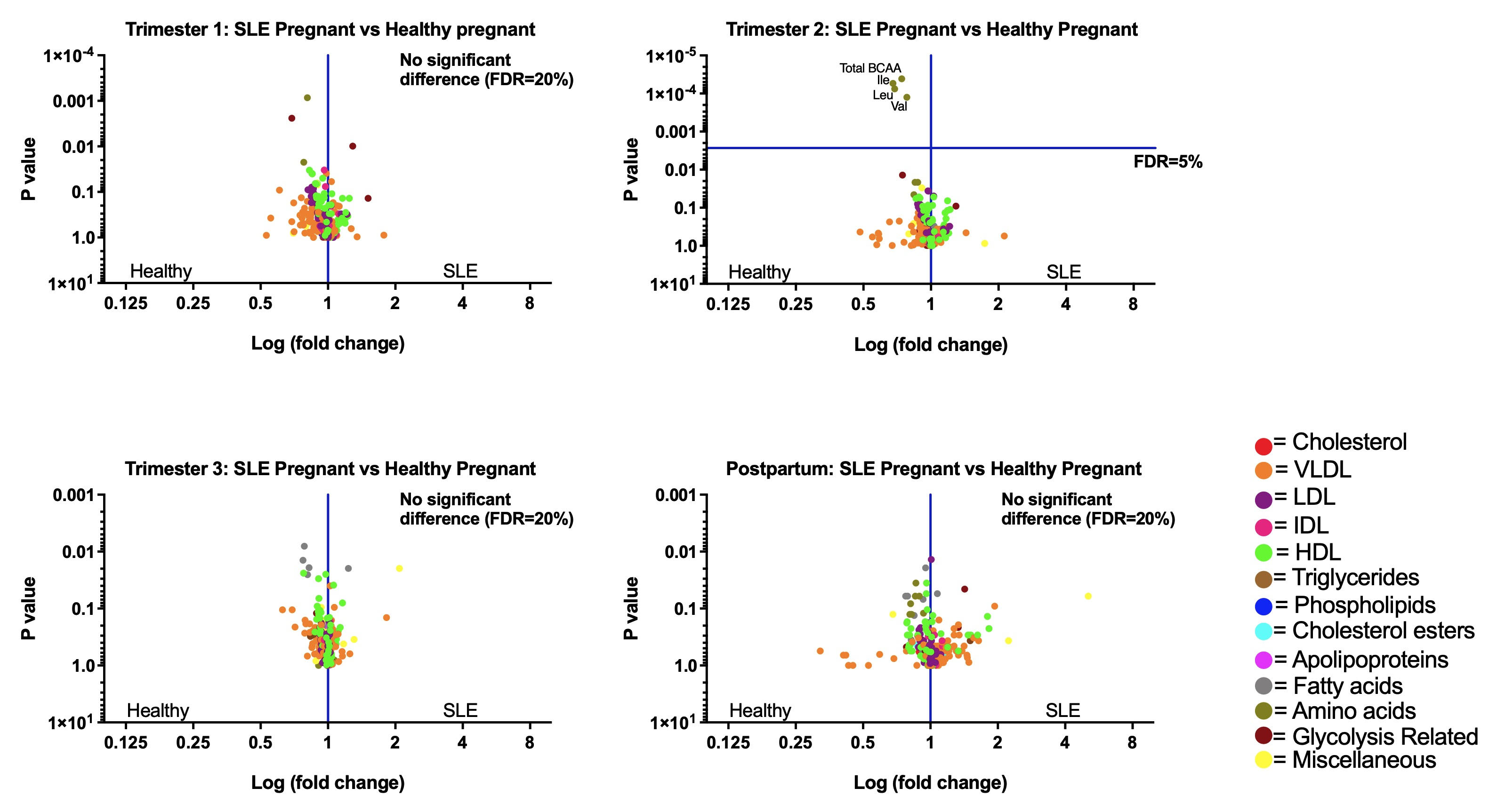
In contrast, SLE and healthy pregnancy revealed a similar anabolic phase in pregnancy, followed by a postpartum catabolic phase, with no significant differences in lipids, amino acids and carbohydrates in the first trimester, third trimester or postpartum. Second trimester serum levels of branch-chained amino acids (isoleucine, valine, leucine, and total branch-chained amino acids) were significantly higher in the healthy pregnant group, compared to the SLE pregnant group (multiple Mann-Whitney tests, FDR=5%, see Figure 2).
Metabolites such as low-density lipoproteins, correlated with disease activity measured using the BILAG (British Isles Lupus Assessment Group)-2004 index in non-pregnant and pregnant SLE groups. Most BILAG correlates were unique to each trimester. Preliminary data indicated that some high density lipoproteins correlated with pre-eclampsia and pregnancy-induced hypertension in SLE pregnancy (Pearson's correlation, FDR=20%).
Conclusion: The distinction between metabolic profiles of SLE and healthy controls found outside of pregnancy was lost during the first and third trimesters of pregnancy and postpartum. Second trimester levels of four branch-chained amino acids were lower in SLE pregnancies compared to healthy pregnancies. Low-density lipoproteins correlated with disease activity in SLE. Further work is needed to understand the use of metabolomics in identifying disease flares and APO in SLE pregnancy.
.jpg) Figure 1: Method chart outlining study groups, collection timepoints, data collected and statistical analysis methods used.
Figure 1: Method chart outlining study groups, collection timepoints, data collected and statistical analysis methods used.
Abbreviations: VLDL: very low density lipoprotein, LDL: low density lipoprotein, IDL: intermediate density lipoprotein, HDL: high density lipoprotein, KNN: K nearest neighbour, T1: first trimester, T2: second trimester, T3: third trimester.
.jpg) Table 1: Demographic and clinical table comparing pregnant SLE and non-pregnant SLE cohorts.
Table 1: Demographic and clinical table comparing pregnant SLE and non-pregnant SLE cohorts.
Abbreviations: BMI: body mass index, ANA: antinuclear antibodies, dsDNA: anti-double stranded DNA antibodies, BILAG: British Isles Lupus Assessment Group.
 Figure 2: Volcano plots demonstrating a similar metabolic pattern in systemic lupus erythematosus (SLE) pregnancy and healthy pregnancy, with most metabolites peaking in the third trimester. P values calculated using multiple Mann-Whitney tests, significance threshold determined via FDR (false discovery rate) as indicated and represented via a horizontal line. Values to the left of the vertical line were higher in the healthy pregnant group and values to the right higher in the SLE pregnant group. Significantly higher levels of total branch-chained amino acids (p= 0.000041), isoleucine (p= 0.00054), leucine (p=0.000075) and valine (p=0.000125) in the second trimester of the healthy pregnant cohort.
Abbreviations: BCAA: branch-chained amino acids, Ile: isoleucine, Leu: leucine, Val: valine, VLDL: very low density lipoprotein, LDL: low density lipoprotein, IDL: intermediate density lipoprotein, HDL: high density lipoprotein, FDR: false discovery rate.
Disclosures: S. Luong, None; I. Sheldon, None; C. Raine, None; E. Jury, None; I. Giles, UCB.
Background/Purpose: Systemic lupus erythematosus (SLE) is an autoimmune disease characterised by activation of immunological and cellular pathways, disease flares and adverse pregnancy outcomes (APO). Metabolomic analysis of metabolites can identify biomarkers of disease activity or APO in SLE. This is the first prospective longitudinal study of pregnant and non-pregnant SLE patients to identify the impact of pregnancy on metabolites and associations with disease activity.
Methods: Non-pregnant SLE, pregnant SLE, and healthy pregnant patients recruited from tertiary hospital clinics, and serum obtained during each trimester and 3-6 months postpartum for pregnant patients, or every 3-6 months for non-pregnant SLE patients from 2018-2020. All SLE patients met ACR classification criteria. Healthy non-pregnant controls recruited from university staff. Serum sent for metabolomic analysis using nuclear magnetic resonance spectroscopy (Figure 1).
Results: 19 non-pregnant SLE (mean age 31yrs), 17 pregnant SLE (mean 34.48yrs) and 26 healthy pregnant (mean 34.84yrs) patients were compared to 45 non-pregnant healthy controls (mean age 32.69yrs). See Table 1 for clinical details of SLE patients. Rates of APO in SLE and healthy pregnant cohorts respectively: miscarriage (17.6% vs 7.7%), stillbirth (0% vs 0%), premature birth (11.8% vs 0%), caesarean section (52.9% vs 5.7%) and pre-eclampsia (5.9% vs 0%).
Differences between non-pregnant SLE and healthy control metabolomes were shown by two binary classification models (decision tree and random forest), with classification accuracies >80% using metabolite measurements and 10-fold cross-validation. Acetoacetate and docosahexaenoic acid were the top metabolites in the decision tree model, with 96% classification accuracy.
In contrast, SLE and healthy pregnancy revealed a similar anabolic phase in pregnancy, followed by a postpartum catabolic phase, with no significant differences in lipids, amino acids and carbohydrates in the first trimester, third trimester or postpartum. Second trimester serum levels of branch-chained amino acids (isoleucine, valine, leucine, and total branch-chained amino acids) were significantly higher in the healthy pregnant group, compared to the SLE pregnant group (multiple Mann-Whitney tests, FDR=5%, see Figure 2).
Metabolites such as low-density lipoproteins, correlated with disease activity measured using the BILAG (British Isles Lupus Assessment Group)-2004 index in non-pregnant and pregnant SLE groups. Most BILAG correlates were unique to each trimester. Preliminary data indicated that some high density lipoproteins correlated with pre-eclampsia and pregnancy-induced hypertension in SLE pregnancy (Pearson's correlation, FDR=20%).
Conclusion: The distinction between metabolic profiles of SLE and healthy controls found outside of pregnancy was lost during the first and third trimesters of pregnancy and postpartum. Second trimester levels of four branch-chained amino acids were lower in SLE pregnancies compared to healthy pregnancies. Low-density lipoproteins correlated with disease activity in SLE. Further work is needed to understand the use of metabolomics in identifying disease flares and APO in SLE pregnancy.
.jpg) Figure 1: Method chart outlining study groups, collection timepoints, data collected and statistical analysis methods used.
Figure 1: Method chart outlining study groups, collection timepoints, data collected and statistical analysis methods used. Abbreviations: VLDL: very low density lipoprotein, LDL: low density lipoprotein, IDL: intermediate density lipoprotein, HDL: high density lipoprotein, KNN: K nearest neighbour, T1: first trimester, T2: second trimester, T3: third trimester.
.jpg) Table 1: Demographic and clinical table comparing pregnant SLE and non-pregnant SLE cohorts.
Table 1: Demographic and clinical table comparing pregnant SLE and non-pregnant SLE cohorts. Abbreviations: BMI: body mass index, ANA: antinuclear antibodies, dsDNA: anti-double stranded DNA antibodies, BILAG: British Isles Lupus Assessment Group.
 Figure 2: Volcano plots demonstrating a similar metabolic pattern in systemic lupus erythematosus (SLE) pregnancy and healthy pregnancy, with most metabolites peaking in the third trimester. P values calculated using multiple Mann-Whitney tests, significance threshold determined via FDR (false discovery rate) as indicated and represented via a horizontal line. Values to the left of the vertical line were higher in the healthy pregnant group and values to the right higher in the SLE pregnant group. Significantly higher levels of total branch-chained amino acids (p= 0.000041), isoleucine (p= 0.00054), leucine (p=0.000075) and valine (p=0.000125) in the second trimester of the healthy pregnant cohort.
Abbreviations: BCAA: branch-chained amino acids, Ile: isoleucine, Leu: leucine, Val: valine, VLDL: very low density lipoprotein, LDL: low density lipoprotein, IDL: intermediate density lipoprotein, HDL: high density lipoprotein, FDR: false discovery rate.
Disclosures: S. Luong, None; I. Sheldon, None; C. Raine, None; E. Jury, None; I. Giles, UCB.

