General IR
The creation of ex vivo models for IR training of medical students and residents.
- JL
Jorge Lopera, MD, FSIR (he/him/his)
Faculty
UT Health Science Center in San AntonioDisclosure information not submitted.

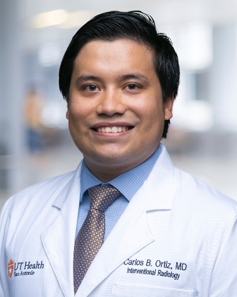
Carlos B. Ortiz, MD
Resident Physician
UT Health San Antonio.jpg)
Marina Borrego, BS
Research Laboratory Technician
UT Health San Antonio- JW
John A. Walker, MD
Associate Professor of Radiology
UT Health SA - Radiology MC7800 - RS
Rajeev Suri, MD
Professor
University of Texas Health San Antonio
Abstract Presenter(s)
Author/Co-author(s)
To report our experience with the development of several teaching models to teach basic and advanced IR techniques using ex- vivo organs.
Background: Simulation in IR is very limited due to the high cost of computer base simulators and the lack of realistic experience when using plastic and 3 D models. We have created successfully several teaching models using ex- vivo organs to teach and train on several basic and advanced IR techniques.
Clinical Findings/Procedure Details:
The following models were developed and tested by different operators with different levels of training:
PTC model: after the biliary system was opacified using gallbladder injection operators performed a PTC with placement of an internal external drain. The model simulates a PTC in mildly dilated biliary system, US punctures of the bile ducts is also possible.
PCN model: Operators developed the skills to perform a PCN using US and fluoroscopic guidance, different levels of hydronephrosis can be simulated injection contrast through a cannulated ureter
Double J ureteral model: Kidneys with long pieces of ureter were used. Operator deployed a DJS using an antegrade access. The model allows to simulate exchange of DJS using a retrograde access.
TIPS and TJLB model: the model allows to perform cannulation of the HV and TJLB, puncture of the portal vein under fluoro or intravascular ultrasound and simulate all the steps of a TIPS with placing a Viatorr stent graft.
Angiographic and Embolization model: Operators can learn basics of DSA, road mapping and selective catheterization of different vessels. A flow model was developed that allows to perform basic embolization techniques using different embolization, the renal artery, the hepatic artery and the portal vein can be used to practice in different vessels sizes and flow situations.
Biopsy and drainages models: Organs can be used to learn basics of liver and kidney biopsy. Percutaneous cholecystostomy tube can be practiced.
Conclusion and/or Teaching Points: Ex- vivo models offer a very economical and realistic simulation for different IR procedures that are very useful to teach basic and advanced techniques to medical students, residents and fellows.

.jpg)
.png)
.jpg)
.png)
.png)
.png)
.jpg)
.png)
.png)