Imaging
Quantitative MRI texture analysis for evaluating treatment response following Irreversible electroporation ablation in hepatocellular carcinoma

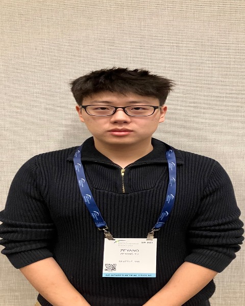
Zeyang Yu, MS (he/him/his)
Research volunteer (master student)
University of WashingtonDisclosure(s): No financial relationships to disclose
- AE
Aydin Eresen, PhD (he/him/his)
Assistant Researcher
University of California, IrvineDisclosure(s): No financial relationships to disclose

Zigeng Zhang, MD
Postdoctoral Fellow
University of California, Irvine- JT
Jiaxin Tan, n/a (he/him/his)
Researcher
Western University - QH
Qiaoming Hou, n/a
Lab Manager
University of California, Irvine 
Farouk Nouizi, PhD
Assistant Researcher
Radiological Sciences - UCI- VY
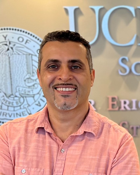
Vahid Yaghmai, n/a
Chairman of Radiology
University of California, Irvine - ZZ
Zhuoli Zhang, PhD
Professor
University of California, Irvine
Presenting Author(s)
Author/Co-author(s)
Contrast-enhanced imaging is used to evaluate therapeutic result of Irreversible electroporation (IRE). However, it requires invasive interaction with patients and limited reproducibility due to varying post-processing approaches. In this study, we suggested using quantitative analysis of tissue texture captured with conventional MRI data as an alternative method for defining the behavior of IRE ablation outcomes for early assessment.
Materials and Methods:
To generalize underlying structural alterations of tissues under various conditions, a total of twenty-six rabbits were designed and used in this study. We implemented IRE ablation to performed on solid liver cancers of eighteen rabbits and normal liver tissues of eight VX2 rabbits, imaging data representing the treatment outcome including anatomical (T1W and T2W) and contrast-enhanced magnetic resonance imaging was acquired with a 3T clinical Siemens scanner. In addition, IRE ablation regions were delineated by the perfusion images, and regions of interest (ROIs) were translated to anatomical MRI data. Through the study, we examined the patterns and heterogeneity in the ROI texture to identify redundant variables and drop them to increase the productivity and overall quality of the data. We established a statistical model that combined the integration of four variables with an optimized SVM framework and chose the five-fold cross-validation as the method of evaluation. The performance of the model was evaluated using the area under the receiver operating characteristics curve.
Results:
Overall, nineteen T1W and twenty T2W MRI features were chosen as candidates for the representation of distinctive changes associated with the IRE ablation procedure after the pairwise interaction of the variables. A statistical model integrating three imaging features from T1W, and T2W MRI was constructed and fitted, through the evaluation process, the model reaches 90% and 85% accuracies for training and validation sets. Additionally, T1W and T2W MRI models produced AUC values of 0.91 and 0.93 for discriminating IRE ablation regions, respectively.
Conclusion:
The results proved that quantitative analysis of tissue texture captured with conventional MRI can be used to describe underlying changes in different tissues following IRE ablation and can serve as noninvasive biomarkers for evaluating treatment response immediately.

.png)
.jpg)
.png)
.png)
.png)
.jpg)
.png)
.png)
.jpg)