038 - The tortuous road less traveled: Complex management of varicose veins in Klippel-Trenaunay syndrome.
John Hufnagle, MD – Resident Physician, Yale School of Medicine; Shin Mei Chan, MS4 – Medical Student, Yale School of Medicine; Todd Schlachter, MD – Assistant Professor, Yale School of Medicine; Jeffrey Pollak, MD – Professor and Director of HHT, Yale School of Medicine; Angelo Marino, DO – Assistant Professor, Yale School of Medicine
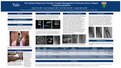
Purpose: Klippel Trenaunay Weber syndrome (KTS) is a congenital triad of capillary malformations, varicose veins, and limb overgrowth. Varicose veins in these patients are often extensive, debilitating and psychologically distressing. This study reviews endovascular varicose vein treatment pearls and pitfalls in KTS patients.
Material and Methods: This study retrospectively reviewed three KTS patients with debilitating and distressing varicose veins, treated at our institution.
Results: Endovascular therapy in KTS patients is made challenging by the extensiveness of varicose veins and the underlying valveless embryological veins. Work-up begins with contrast enhanced MRI and reflux ultrasound to evaluate for the presence of embryological veins, patency and caliber of the deep venous system and the valvular competence of the superficial venous system. A lateral marginal vein was the primary culprit of reflux in two cases, with an accessory great saphenous vein in the other. Prior to treatment, ultrasound and venography were used to confirm that the deep veins were patent.
Endovascular treatment first targets the embryological veins, followed by occlusion of incompetent superficial veins. Common options include endovenous ablation, sclerotherapy, coil embolization and ambulatory phlebectomy. In our institution, embryological veins were first treated with endovenous ablation, followed by phlebectomy/sclerotherapy of the varicose and incompetent perforating veins. In one patient, the lateral marginal vein was >12mm in diameter and following endovenous ablation, complete occlusion was subsequently achieved with coil embolization. Varicosities can be exacerbated if the great saphenous vein is occluded prior to treatment of the lateral marginal vein. This was the case in two of the patients, for which a total of six and fifteen treatment sessions were required. Significant clinical and cosmetic improvement was achieved in all cases. Recurrence invariably occurs; however, this can be retreated with the same regimen. In one case, temporary malleolar skin ulceration occurred.
Conclusions: Varicose veins in KTS patients can be effectively treated with endovascular therapy, although several treatments are required, and recurrence should be expected. Prior to treatment, it is important to evaluate the vascular anatomy, avoid removing a functioning great saphenous vein and ensure that the deep venous system is patent.