065 - MRI Radiomics Correlate with Clinical Outcomes of Peripheral Venous Malformations Following Percutaneous Sclerotherapy
Purpose: To study the changes in MRI radiomic features occurring in peripheral venous malformations (VMs) after percutaneous sclerotherapy.
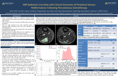
Material and Methods: Our vascular anomalies database was searched for patients with extremity VMs who were treated by image-guided percutaneous sclerotherapy, had completed their treatment plan, and clinical and imaging follow-up between 2005-2022. VMs were manually segmented on pre- and post-treatment T2-WI MRI using 3D Slicer software to assess the changes in lesion volume and signal intensity (SI). To account for signal parameter differences between pre- and post-treatment T2-WI, the post-treatment T2-WI signal was re-scaled to the pre-treatment T2-WI using a simple histogram matching algorithm, which allowed for precise and true calculation of SI change. Therapeutic response was categorized as 0=worse or unchanged and 1=improvement based on clinical evolution after treatment. Clinical outcome assessment was also scored on a 7-point scale, from -3 (worst) to +3 (maximum improvement), based on patient perception of symptom improvement. Spearman's rank correlation coefficient (ρ) and Paired t-test (t) were used for statistical analysis.
Results: Eighty-one patients (mean age: 20±14 years; 47 females) with peripheral upper (23 lesions) and lower (58) extremity VMs underwent 125 percutaneous sclerotherapy (range:1-6). Different sclerosants were used: alcohol (52 sessions), bleomycin (38), and sotradecol (35). Most patients (77) reported clinical improvement following sclerotherapy, including mild (8 patients), moderate (22), and significant (47) improvement. The mean change in lesion volume was -7.9±24.6 cm3 (P=.005) and in mean SI was -123.1±162.9 (P <.001). Overall, there was a significant correlation between the change in lesion volume and treatment response (ρ=-.3, P=.004). On subgroup analysis, SI change correlated with clinical outcomes in patients only receiving one treatment (n=51; ρ=-.3, P=.01) and lesions treated with bleomycin (n=22; ρ=-.4, P=.04). Lesion volume change correlated with clinical outcomes of pediatric patients (n=50; ρ=-.3, P=.03), lesions treated with sotradecol (n=17; ρ=-.5, P=.02) and lesions located in foot (n=10; ρ=-.6, P=.04).
Conclusions: MRI radiomic features, including lesion volume and signal intensity, correlate with the clinical outcomes of peripheral venous malformations treated using percutaneous sclerotherapy.