040 - Pulmonary Thomboembolus: What Electron Microscopy Teaches Us
Vishal Arora, M.D. – Professor, Cardiologist, Internal Medicine, Cardiology, Medical College of Georgia; Islam Elhelf, M.D. – Assistant Professor, Interventional Radiology, Diagnostic Radiology, Medical College of Georgia; Rimaz Khadir, B.S. – Medical Student, Medical College of Georgia; Brendan Marshall, PHd – Professor and Director of Electron Microscopy, Cell Biology, Medical College of Georgia; Musa Sharkawi, MBBSCh. – Asssistant Professor, Cardiology, Medicine, Cardiology, Medical College of Georgia; Hanping Wu, M.D. – Assistant Professor, Interventional Radiology, Diagnostic Radiology, Medical College of Georgia
Purpose: This investigation was undertaken to investigate the composition of fibrin in pulmonary embolectomy thomboemboli specimens, after the observation that most emboli contain portions of older, tan-white appearing firm, rubbery thrombus in addition to recent soft, dark red thrombus, retrieved using the Inari FLowTriever device.
Material and Methods: Thrombus obtained using the FlowTriever was visually examined and the most chronic appearing portions were selected for electron microscopy (EM). Images were reviewed with the electron microscopist and findings for each sample were recorded along with representative photomicrographs. In most cases a photograph of the gross specimen was also reviewed.
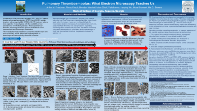
Results: In over 50% of patients there were chronic appearing changes in at least a portion of the gross thrombus. EM of older appearing thrombus demonstrated the following: Thick, frayed-end broken fibrin strands, dense sheets of thick fibrin with scant cellularity, extensive leukocyte infiltration and phagocytosis indicating attempted resolution through the inflammatory system, liposomes and neutrophil extracellular traps (NETS), suggesting marked chronicity. Recent thrombus instead showed relatively sparse long thin intact fibrin strands and numerous red blood cells.
Conclusions: Profound differences exist between fresh new thrombus thin fibrin strands and older, dense, thickened fibrin, frequently coexisting with dense leukocytes indicating inflammation. These findings suggest a reason for insoluble older thrombus using exogenous plasminogen activators, which otherwise effectively resolve fresh, more recent emboli. The presence of organized thrombus with dense sheets of thick fibrin and white blood cells, which would be expected to resist plasmin and may correlate with the prevalence of chronic thromboembolic pulmonary hypertension (CTEPH) and long-term pulmonary disability seen in 4-6% and 30-43% of PE patients respectively. As this investigation continues we hope to provide some framework of understanding of the resolution or lack thereof in pulmonary emboli. Could the current PE management algorithms be optimized towards embolectomy in lieu of thrombolysis or even anticoagulation alone in some patients? Furthermore, it is hoped that this study will propagate interest in correlation with CTA imaging findings to predict which patients are more likely to require embolectomy, rather than thrombolytic or anticoagulation only treatments.