092 - 3D/4D Vessel Cast in the Evaluation of Carotid Artery Stenosis
Rafey Khan, BS – Medical Student, Florida International University Herbert Wertheim College of Medicine; Collin Shick, BS – Medical Student, University of North Carolina School of Medicine; Sam Salazar, BS – Medical Student, Florida International University Herbert Wertheim College of Medicine; Austin Pourmoussa, MD – Resident, Radiology, Mount Sinai Medical Center; Muhammad Hasan, MBBCh, RPVI, RVT – Cardiovascular Clinical Education and Accreditation, Miami Cardiac & Vascular Institute; Brian Schiro, MD, FSIR, RPVI – Interventional Radiologist, Miami Cardiac & Vascular Institute
Purpose: Screening for carotid artery stenosis (CAS) is critical in symptomatic patients as stenosis related to atherosclerotic plaque can cause neurological ischemia. Current first-line evaluation for CAS is carotid duplex ultrasonography (DUS); however, precise determination of stenosis is limited. The reference standard for determining the exact severity of CAS is a catheter angiogram (CA). Though very effective, CA is invasive and thus only used in select patients, primarily those who are suspected to need treatment. 3D/4D vessel cast is an ultrasound technique that provides an accurate map of the luminal surface of the carotid artery. The purpose of this study is to validate 3D/4D vessel cast as a useful tool in the evaluation of CAS and compare its accuracy to CA and carotid DUS.
Material and Methods: This single-center, retrospective cohort study comprised of 18 events among 14 patients with CAS from 2018 to 2022. Patients with a carotid DUS, associated 3D/4D vessel cast, and a carotid CA within a 1-year time frame were included. All vessel casts were obtained with the XL14-3 workflow (Philips, Amsterdam, Netherlands). Measurements of the stenotic vessel were obtained on the vessel cast image and compared to values from the CA and DUS reports. The primary endpoint was the difference in percent stenosis between the vessel cast and CA. Additionally, a binary analysis comparing reported stenosis of the vessel cast/CA to the reported range of DUS was performed.
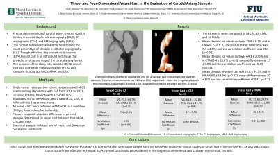
Results: Of the 18 events, 3D/4D vessel cast and carotid CA yielded an average percent stenosis of 73.9 ± 0.06% and 77.8 ± 0.10% respectively. The average difference between the groups was 7.4 ± 0.05%. There was no significant difference in calculated percent stenosis between imaging modalities (p=0.06). The two techniques yielded a correlation coefficient of 0.57 (p = 0.013), demonstrating moderate strength. When compared to the reported range of stenosis from the DUS, it was found that vessel cast was in range in 38.9% events (7/18) and CA was in range in 50% of events (7/19). This comparison was not statistically significant (p=0.74).
Conclusions: 3D/4D vessel cast demonstrates moderate correlation to carotid CA in the evaluation of CAS. Further studies are needed to determine the clinical utility of vessel cast. Given that it is both a safe and effective imaging modality, it should be considered in the diagnostic armamentarium to obtain accurate estimates of stenosis.