113 - Optimal Energy Delivery of Auryon Laser to Disrupt Medial Calcification in Tibial Arteries: Cadaveric Study
Purpose: To evaluate the impact of the Auryon Laser Atherectomy system in heavily calcified human cadaver peripheral arteries using microCT.
Material and Methods: Four human limbs were screened via angiography for stenotic and heavily calcified lesions had the arteries dissected for experimentation. Prior to treatment, each vessel was imaged using microCT to establish a baseline. We treated 2-4 segments with Auryon laser per major vessel in the above the knee (superficial femoral/popliteal) (SFA) and below the knee (anterior and/or posterior tibial) arteries (ATA, PTA). We treated 2-4 segments in each vessel with balloon angioplasty as our control. Different laser parameters were used and decided before treatment to explore different treatment effects based on number of passes, and laser energy. After treatment each vessel segment was reimaged using microCT to observe treatment effects.
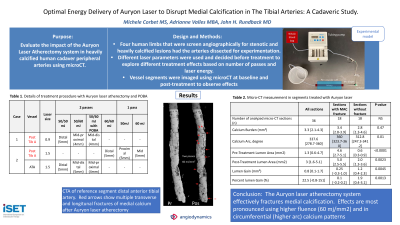
Results: In this ex-vivo model, longitudinal and transverse fractures of medial vessel wall calcification was observed in all tibial specimens containing plate-type (circumferential non-interrupted) patterns calcification and when treated with 60 mJ/mm2 using 0.9 and 1.5 mm sized lasers. This effect was consistently noted when higher laser energy was delivered either alone or following lower energy delivery. A single laser pass using smaller laser catheters 50 mJ/mm2 didn't impact medial calcium although sampling was limited, with only 2 segments containing medial plate-type calcification were treated with this energy delivery algorithm alone. Notably, medial plate calcification was unaffected in control tibial samples treated with angioplasty alone. No definitive impact was seen with in the larger SFA specimens when 2.0 mm lasers were utilized; there was no evidence of intimal calcification disruption in any specimens. Sections with calcium cracks had a significantly greater degree of calcification arc as compared to the sections without cracks (343 ± 28 vs. 264 ± 106 degrees, p=0.0011), the calcium burden was not different (3.8 ± 1.1 vs. 3.8 ± 2.8, p=0.89). No acute occlusion or no transmural dissection was observed on histologic evaluation.
Conclusions: This exploratory study provides evidence that the Auryon laser is capable of fracturing medial arterial calcification through a secondary mechanism of action, photomechanical shockwaves. Further work is necessary to define frequency and limitations of this phenomenon.