015 - Non-Fluoroscopic 3D Image Guidance for PAD Interventions: An Initial Preclinical Study in Five Vascular Phantoms
Purpose: Assess the potential role of utilizing a novel 3D electromagnetic navigational system as an adjunct to conventional 2D fluoroscopy for patients with peripheral arterial disease (PAD).
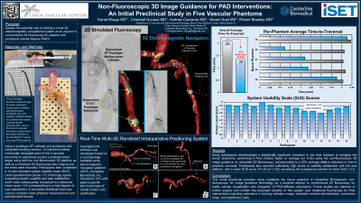
Material and Methods: Five 3D-printed bench phantoms designed to mimic tortuous multifocal calcified PAD were created, based on source CT datasets from real patients. Investigational software was developed based on a commercially-available aortic electromagnetic navigation platform (IOPS, Centerline Biomedical, Cleveland, OH), with patient-specific structural maps of vessel lumens and calcification. Using a prototype 6Fr catheter and guidewire with integrated tracking sensors, 15 interventionalists individually traversed the length of all 5 vascular phantoms using both the non-fluoroscopic 3D platform as well as a simulated 2D fluoroscopy-like imaging and the times were recorded. Participants then completed a 10-item standard system usability scale (SUS) Likert questionnaire (score 1-5, 5=strongly agree) evaluating system usability and user satisfaction, which were subsequently compared to a reference mean score >3.5 corresponding to a high degree of user satisfaction. A one-tailed statistical t-test was used to compare mean phantom traversal times and questionnaire scores.
Results: Study participants demonstrated a statistically significant reduction in the time required to navigate the bench phantoms, performing 0.7min (42sec) faster on average (p < 0.05) using the non-fluoroscopic 3D image guidance when compared with simulated 2D fluoroscopy, corresponding to a 25% average relative reduction in time to phantom traversal. Participants also reported sufficiently high levels of usability satisfaction with the new platform, with a mean SUS score of 4.29 (p < 0.05), exceeding the acceptance criterion.
Conclusions: This small preclinical phantom study highlights the future potential of Centerline Biomedical’s non-fluoroscopic 3D image guidance technology as a possible adjunct to conventional 2D fluoroscopy for highly precise visualization and navigation of PAD-afflicted vasculature. Future studies are planned to further explore and confirm the proposed benefits of this system over traditional fluoroscopy for PAD interventions including reductions in ionizing radiation usage, iodinated contrast administration, procedure times, and healthcare costs.