Back

Women’s Health
083 - Presence of Cervicovaginal Arterial Branches in Uterine Fibroid Embolization (UFE): Clinical Significance and Outcomes

Rohit Reddy, BSc – Research Fellow, Interventional Radiology, University of Miami; Kenneth Briley, MD, PhD – Fellow, Interventional Radiology, University of Miami; Arais Cavada, MSN – Nurse Practitioner, Interventional Radiology, University of Miami; Shree Venkat, MD – Associate Professor, Interventional Radiology, University of Miami; Zoe Miller, MD – Associate Professor, Interventional Radiology, University of Miami; Shivank Bhatia, MD – Professor & Chair, Interventional Radiology, University of Miami; Prasoon Mohan, MD – Associate Professor, Interventional Radiology, University of Miami
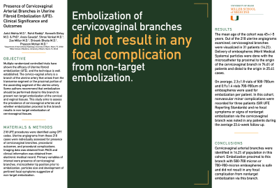
Purpose: Multiple randomized controlled trials have shown the efficacy of Uterine fibroid embolization (UFE), and the technique is well established. The cervico-vaginal artery is a branch of the uterine artery that arises from the transverse segment or the proximal portion of the ascending segment of the uterine artery. Some authors recommend that embolization should be performed distal to this branch to prevent non target embolization of the cervical and vaginal tissues. This study aims to assess the prevalence of cervicovaginal arteries and whether embolization proximal to this branch results in non-target embolization of cervicovaginal tissues.
Material and Methods: 218 UFE procedures were identified using CPT codes. Uterine angiograms from these 218 cases were individually assessed for presence of cervicovaginal branches, procedural outcomes, and procedural complications. Imaging data was obtained from PACS and clinical information was obtained from electronic medical record. Primary variables of interest were presence of cervicovaginal branches, microcatheter tip position prior to embolization, particles size and development of pertinent focal symptoms suggestive of non-target embolization.
Results: The mean age of the cohort was 45+/-5 years. Out of the 218 uterine angiograms examined, cervicovaginal branches were visualized in 31 patients (14.2%). Delivery of embospheres (Merit Medical Systems) particles were done with the microcatheter tip proximal to the origin of the cervicovaginal branch in 74.6% of patients and distal to the origin in 25.4% cases. On average, 2.3±1.8 vials of 500-700um and 0.9±1.4 vials 700-900um of embospheres were used for embolization per patient. In this cohort, nonvascular minor complications were recorded for three patients (SIR UFE Reporting Standards) and no focal symptoms or signs of nontarget embolization via the cervicovaginal branch was noted in any patients during the average 23.4-week follow up.
Conclusions: Cervicovaginal arterial branches were identified in 14.2% of population in this cohort. Embolization proximal to this branch with 500-700 micron or 700–900-micron embospheres is safe and did not result in any focal complication from nontarget embolization via this branch.
