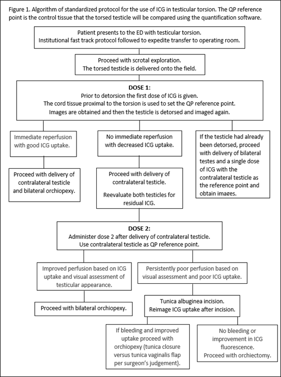
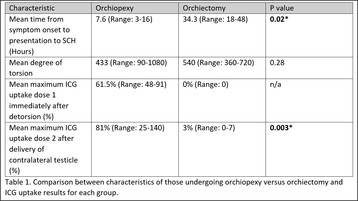
Introduction: The current standard of care to determine testicular viability in testicular torsion is surgeon’s subjective assessment. Studies on objective means to assess testicular perfusion after detorsion are lacking. Indocyanine green (ICG) is a fluorescent dye which has been widely used to evaluate tissue perfusion. To date use of ICG in testicular torsion has been limited to case reports with no series or protocols yet published. We aimed to develop and implement a standardized protocol for the use of ICG in testicular torsion and evaluate the effectiveness of ICG as an objective tool to assess testicular perfusion. Methods: This was a single institution pilot study on the use of ICG in testicular torsion. A useable standardized protocol for use of ICG in testicular torsion was developed and implemented. The Stryker SPY PHI system and novel quantification software, SPY-QP, were used for ICG imaging. The protocol development was an adaptive process. The finalized protocol is detailed in figure one. Results: ICG was used in 17 cases of testicular torsion with 13 undergoing orchiopexy and 4 undergoing orchiectomy. Of the 4 testes which were removed, we noted no to very low ICG uptake with SPY-QP measurements of 0-7% when compared to a mean ICG uptake in the orchiopexy group of 74.5% (p= 0.003) (Table 1). The pathology of all four showed hemorrhagic necrosis. At early post operative follow up there was no evidence of testicular atrophy in those that underwent orchiopexy. There were no intraoperative complications or adverse reactions to ICG. Conclusions: Our institution successfully developed and implemented a standardized protocol for ICG use in testicular torsion. Early results indicate our protocol provides objective and quantitative assessment of testicular perfusion. We think this technology is a useful adjunct in the surgical management of testicular torsion and can aid in surgical decision making particularly in equivocal cases. SOURCE OF Funding: None