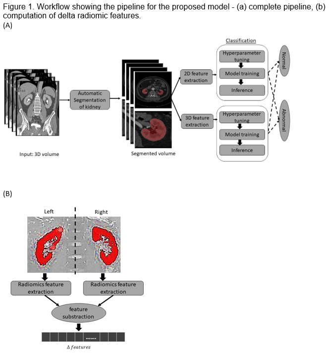
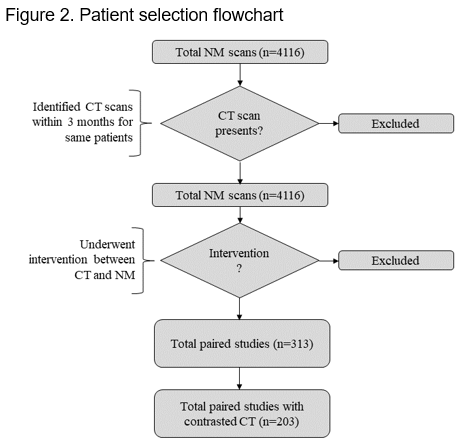
Introduction: Differential kidney function plays an important role in the management of kidney disorders. We sought to assess if this can be obtained through an automated evaluation of contrast enhanced computed tomography using a combination of deep learning and machine learning algorithms without nuclear medical imaging. Methods: All patients who underwent kidney nuclear scanning at Mayo Clinic between 2018-2022 were collected. Computerized tomographic (CT) scans of the kidneys that were obtained within a three-month interval before or after the nuclear scans were extracted. Patients who underwent a urological or radiological intervention within this time frame were excluded. A deep learning segmentation model was used to automatically segment kidneys (Figure 1). 2D and 3D radiomics features were extracted and compared between the two kidneys to compute delta radiomics and assess its ability to predict differential kidney function. The performance of these models was reported using receiver operating characteristics, precision, recall, and F1 score. Results: There were 4118 nuclear medical scans. We randomly selected 303 CT scans for review. Following exclusion, there were 203 paired studies (Figure 2). The fine-tuned segmentation model demonstrated a Sorensen-Dice score of 0.7. The best results were obtained when the support vector classifier model was used on 3D delta radiomics features computed from the venous phase of contrast-enhanced CT scans. This model has an AUC of 0.90 with a precision, recall, and F1 score of 0.80, 0.80, and 0.82 respectively, to detect abnormal kidney function. Conclusions: This proposed automated pipeline can provide important differential kidney function information from contrast-enhanced cross section imaging without the need for dedicated nuclear scans. SOURCE OF Funding: None