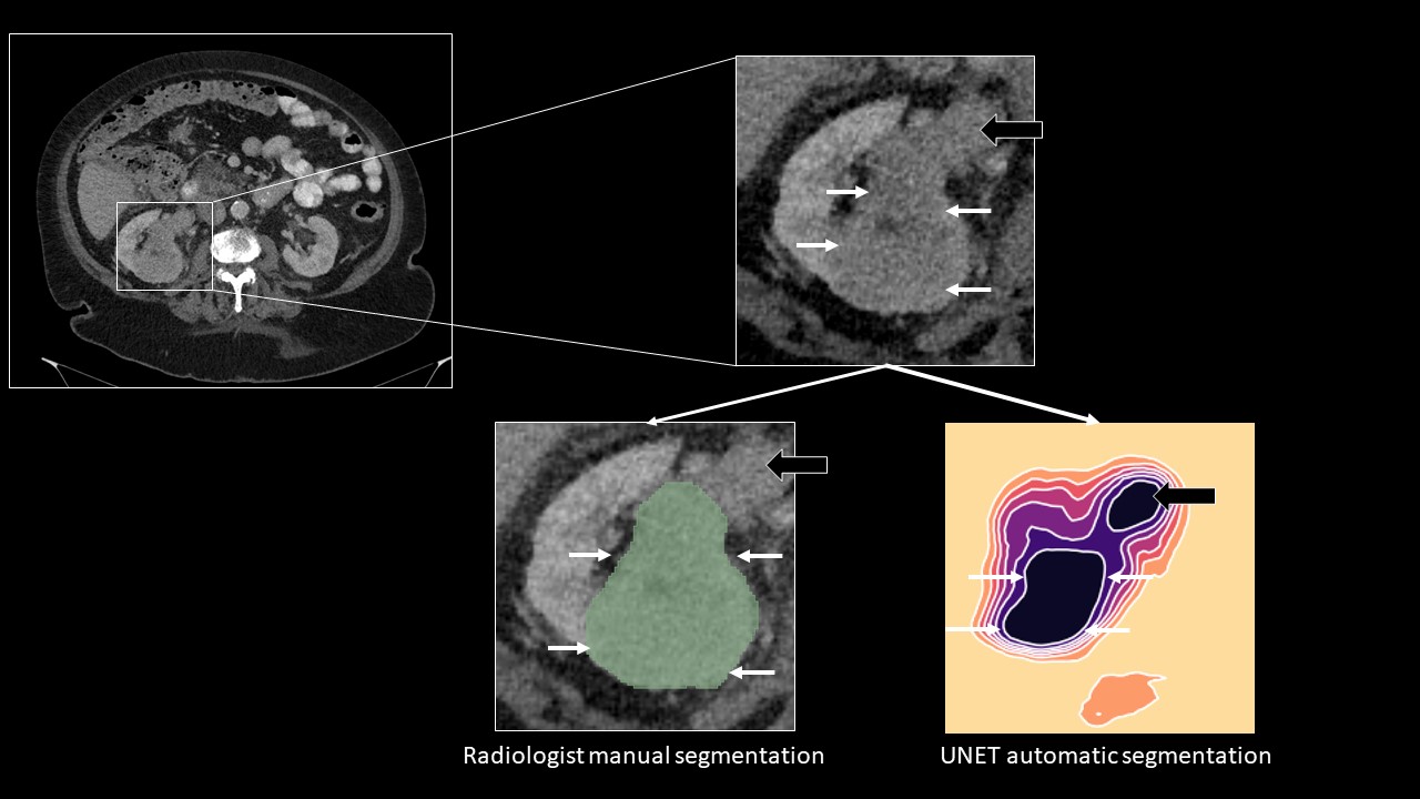
Introduction: Automatic segmentation of renal tumors is a cornerstone for Artificial Intelligence (AI) based tumor diagnostics. This study presents an automatic segmentation algorithm and visualization method using CT-studies from renal tumor patients recruited in a multicenter setting. Methods: Renal tumor patients diagnosed between 2018 and 2021 were retrospectively assessed for this study. Inclusion criteria were CT-imaging of renal tumors in corticomedullary (CM) or nephrogenic (NG) contrast media phase. Patients aged <18years, and those with cystic or infiltrative renal tumors (i.e. lymphomas) were excluded. Manual segmentation of the renal tumors was performed by a GU-radiologist on all axial CT slices. A convolutional neural network (UNET) was trained based on the radiologists´ manual segmentations. In an independent validation dataset, the accuracy of the UNETs predictions of renal tumor contours was compared to the references standard of manual segmentations and quantified using the DICE score. Results: A total of n=394/ n=350 patients with renal tumors imaged in CM /NG phase, respectively, were included (median age 66y; 35% female; median tumor diameter 5.4cm). CT-studies from >20 radiological imaging centers were included with different imaging protocols and slice thickness. The UNET was trained on n=316 CM and n=294 NG contrast phase patients (n=7019 / n=6859 separate CT images). In the independent validation dataset (n=78 / n=56 patients with 1713 / 1298 CT images), the UNET achieved a DICE score of 0.88 and 0.90 for the corticomedullary and nephrogenic CM phase, respectively. The UNET predictions were visualized using a tile-based approach with color-coding and contour-lines that could be overlaid on CT-images to depict varying levels of prediction confidence. Conclusions: A UNET AI-approach yields a robust automatic delineation of renal tumors on CT-images acquired in clinical routine, irrespective of the contrast media phase. Using color-coding and contour-lines that could be overlaid on original CT-images provides an explainable approach to the UNETs predictions and might improve AI-acceptance in clinical practice. SOURCE OF Funding: This study received funding by the 2022 ESR Research Seed Grant. The 2022 ESR Research Seed Grants were kindly supported by an unrestricted, non-exclusive grant from GE Healthcare.