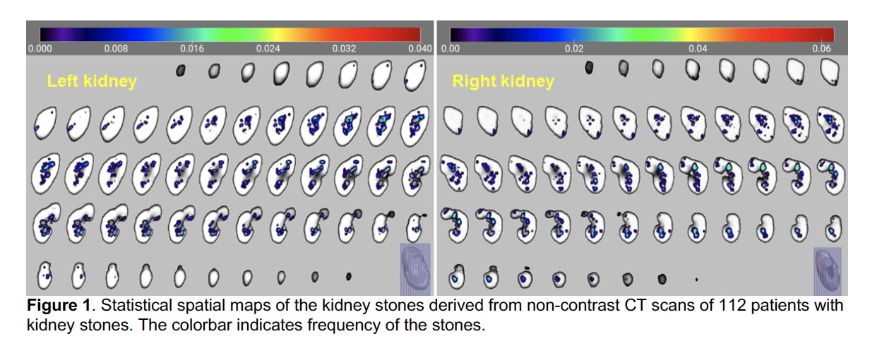
Introduction: Kidney stones vary in size and location. Precise characterization of the kidney stones’ size and location may facilitate effective treatment planning, and spatial mapping of the kidney stones of a group of patients offers a quantitative means to understand how kidney stones spatially distribute across subjects within the kidney. This study evaluated the feasibility to create a kidney atlas, assess individual kidney stones’ location and size, and statistically map kidney stones of a group of patients based on their CT scans using image segmentation and registration algorithms. Methods: Non-contrast CT scans of 122 patients with kidney stones were retrospectively identified. A deep learning (DL) image segmentation model was built to segment kidneys automatically. Kidney stones were automatically identified as outlier voxels with Hounsfield Unit (HU) larger than mean plus 5-sigma of HUs of all voxels within the kidney, and a kidney stone image was generated to have unit intensity value at locations of stone voxels and zero otherwise. Bilateral kidney atlases were created by aligning all kidney images to a manually selected one using affine-transformation and diffeomorphic deformable image registration algorithms subsequently. The kidney stone image of each kidney was spatially transformed to the corresponding left or right kidney atlas with the same image deformation information obtained for creating the atlases. Statistical spatial maps of the kidney stones were generated for the bilateral kidneys separately to quantify frequency of the stones at every voxel of the kidney atlases. Results: The DL image segmentation model segmented the kidneys with an average dice value of 0.96. The automatic stone identification algorithm detected individual kidney stones with 100% sensitivity. Statistical spatial maps of the kidney stones quantified spatial frequency of the kidney stones across subjects, with the highest frequency up to 6%. Conclusions: The statistical spatial mapping of kidney stones provide a quantitative means to characterize frequency of stones within the kidneys. The method can be used to assess kidney stones’ location for individual subjects with reference to group atlases that encode statistical spatial distribution information at a group level. SOURCE OF Funding: NIDDK P20 CHOP/ Penn Center for Machine Learning in Urology (P20DK127488) AUA Care Foundation and SPU Sushil Lacy Research Scholar Award (KMF)