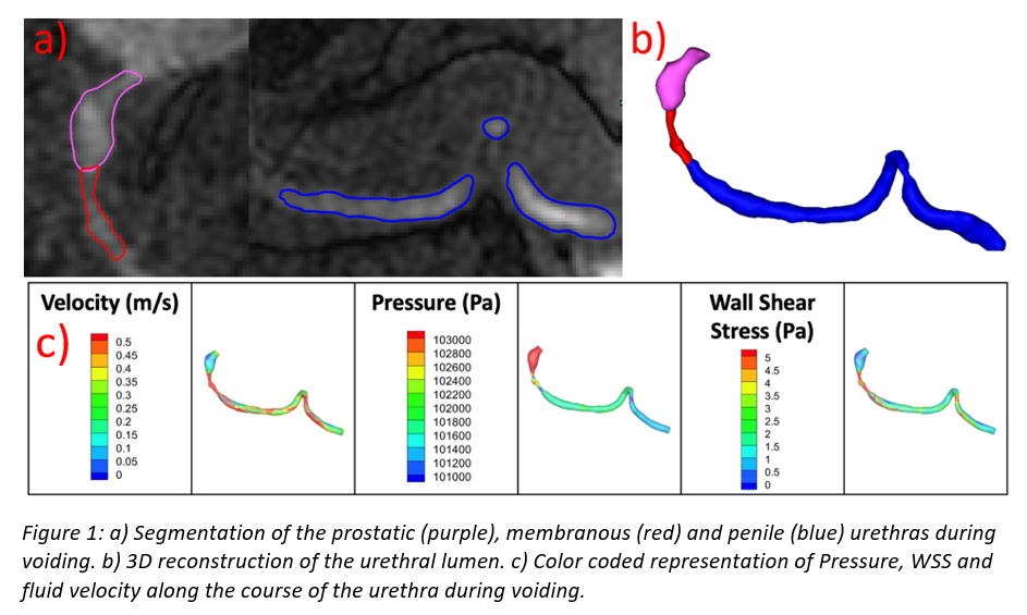
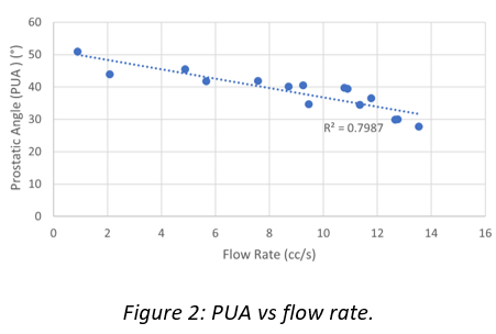
Introduction: Dynamic changes in urethral anatomy during voiding have never been quantitatively assessed. We used real-time magnetic resonance imaging (MRI) to obtain dynamic, high-fidelity 3D images of the lower urinary tract during voiding. These were then used to create accurate real-time urethral reconstructions throughput the voiding event coupled with determinations of urine flow, pressure and wall shear stress by computational fluid dynamics (CFD). Methods: MRI scans were performed on a healthy young man using a clinical 3T scanner according to an IRB-approved protocol to acquire a 3D image every 3.7 s during voiding. 3D models of the urethral lumen were created using MIMICS (Materialise, Leuven, Belgium). Results: Diameters of the internal and external urethral sphincter (IUS, EUS) and prostatic urethral angle (PUA) were obtained throughout voiding. CFD, using computational software to model and simulate fluid behavior under specified constraints of geometry and flow, was used to determine wall shear stress (WSS), pressures and fluid velocities along the length of the urethra during maximum flow (Figure 1). IUS diameter remains unchanged throughout voiding whereas EUS diameter correlates positively with urine flow rate (data not shown here). Most notably, we noted a striking change in PUA during voiding. PUA becomes less acute as flow increases, being straightest at peak flow rate (Figure 2). Conclusions: Real-time MRI can be used to define the biomechanics of the urethra during voiding and reveals – for the first time – a change in prostatic urethral angle during voiding that correlates with urinary flow rate. SOURCE OF Funding: NIH: R01 DK126850-01