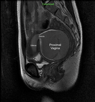
Introduction: Distal vaginal atresia is a unique Mullerian pathology where the urogenital sinus, responsible for the lower 1/3 of the vagina, fails to cannulate during development. Unlike Meyer-Rokitansky-Kuster-Hauser syndrome, which is characterized by agenesis of the upper vagina and uterus, patients with distal vaginal atresia have normal Mullerian development including the uterus and proximal vagina. Methods: We present the case of a 12yo female with a three month history of cyclical abdominal pain. Upon outside workup, she was found to have a short, blind ending vagina. MRI showed a 9cm x 8cm dilated uterus, open cervix and proximal vagina filled with blood products. She was taken to the operating room for combined perineal and robotic transperitoneal pull-through. Results: An 8.5mm camera port was placed in the umbilicus and three 8.5mm working ports were placed using a hidden incision approach below the belt line. We incised the peritoneum and dissected as distally as feasible along the rectovaginal space. At the blind-ending introitus, a U-shaped incision was made to create an anterior flap. This space was dissected proximally, monitoring the correct plane via a finger in the rectum and with direct visualization with the robotic camera. The two planes were connected. A 0-silk was robotically placed to manipulate the vagina. As the vagina was severely dilated from menstrual products, mobility was insufficient to reach the introitus. A small posterior incision on the proximal vagina was made and blood products were partially evacuated. The vesicovaginal space was then dissected robotically, maintaining the lateral attachments as blood supply and avoiding ureteral injury. The vaginal apex was then pulled to the introitus and anastomosed to the anteriorly based flap from the introitus. The patient remained hospitalized for two days with vaginal packing in place. She was trained in the use of vaginal dilators and has been progressively increasing the size without difficulty. Conclusions: Robotic dissection of the proximal vagina in the setting of distal vaginal atresia using a hidden incision approach allows for orthotopic reconstruction, and superior aesthetics as compared to traditional port placement. This approach obviated the need for the use of tissue grafting or more extensive open dissection. SOURCE OF Funding: N/A