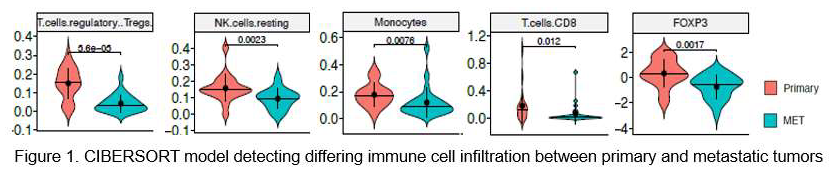
Introduction: Clear cell renal cell carcinoma (ccRCC) is a highly immune infiltrated cancer, but how the tumor immune microenvironment (TIME) evolves during disease progression is unknown. Insights into heterogeneity within primary ccRCC raise questions about potential heterogeneity between primary and paired metastatic tumors. This information may improve understanding of immune evasion leading to metastatic disease and response to immunotherapy. In this study, we characterize and compare the TIME of primary ccRCC with paired asynchronous metastases. Methods: Analysis of ccRCC patients who developed recurrence post radical nephrectomy and had both primary and metastatic tissue available. Whole-transcriptome sequencing was performed on formalin-fixed paraffin-embedded (FFPE) specimens using the illumina platform. Differential expression gene (DEG) analysis was performed using R package edgeR. Gene set enrichment analyses (GSEA) to identify hallmark pathway enrichment was performed using R package fgsea. TIME deconvolution was quantified using CIBEROSRT, an in silico flow cytometry tool. Results: 42 tumor samples from 19 patients (19 primary tumors with 23 matched metastases) were analyzed. Metastasis sites included lung (n=6), bone (n=6), adrenal (n=4), liver (n=2), lymph node (n= 2), and soft tissue (n=3). Angiogenesis and epithelial to mesenchymal transition (EMT) were the most significantly enriched pathways in metastases compared to primary tumors (FDR < 5%). The primary tumors displayed a more immunosuppressive TIME than their matched metastases when comparing immune cell types. (Figure 1). The higher proportion of T regulatory cells (Tregs) in primary tumors was consistent compared against all metastasis tissue types. Metastasis samples displayed higher proportion of M2 macrophages (p=0.003), with the highest proportion in bone and soft tissue. Bone metastases also appeared to be more immunosuppressive in their TIME when compared to lung metastases, with greater Tregs and a lower immune content score. Conclusions: We demonstrate higher infiltration of immunosuppressive cells in the TIME of primary ccRCC compared with metastatic sites. Further studies are necessary to determine the prognostic and therapeutic significance of these findings. SOURCE OF Funding: None