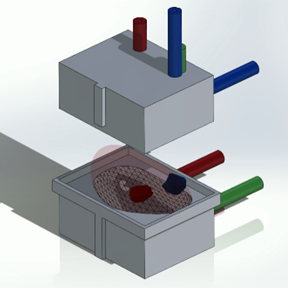
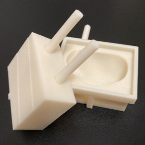
Introduction: Obtaining fresh, viable prostate cancer tissue in a clinical pathology setting is difficult due to lack of optically visible contrast between benign and cancerous tissue. Building on prior methods used to aid correlation of MRI results and whole-mount pathology findings, we have developed an inexpensive and easy to implement means with which 3D-printed molds can be created for any prostate to enable targeted ex-vivo sampling of biopsy-proven cancer lesions seen on MRI. The potential applications for a platform that can reliably sample living prostate cancer tissue are numerous, including the ability to facilitate future cancer research. Methods: STL files of the prostate boundary and target lesions are exported from the UroNav fusion biopsy system. A custom macro was developed for use with SolidWorks (Dassault Systèmes, France), a computer-aided design application. This creates a material-optimized mold around the prostate with needle guides that allow for targeted sampling of up to 4 regions of interest. The guides are customizable in terms of location and dimensions to accommodate different biopsy needles. The parts are then exported and 3D-printed. Following prostate extraction at time of radical prostatectomy, the seminal vesicles are removed. The prostate is then placed into the mold, and samples are taken by inserting a biopsy needle into a guide up to the predetermined depth. The prostate is then passed to pathology for standard processing, while the live tissue in the biopsy cores is available for a variety of purposes. Results: The resulting platform allows for a prostate of any size or morphology to be precisely sampled as long as there is a pre-acquired MRI dataset. The prostate mold allows for accurate sampling of target cancer lesions and is optimized in size for each prostate to allow for minimal material waste. Conclusions: This novel platform provides the unique ability to obtain fresh prostate cancer tissue. The utility of this is not necessarily limited to prostate cancer alone but may be of use in other solid-organ malignancies with targetable lesions as well. SOURCE OF Funding: None