CRTA Post-Doctoral Research Fellow, Urologic Oncology Branch, National Cancer In
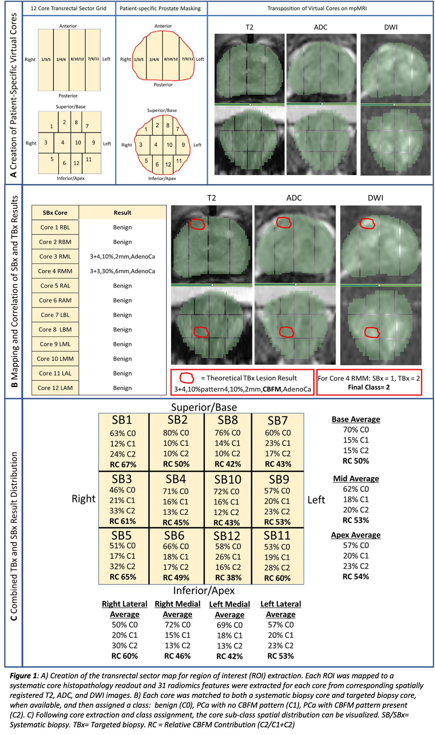
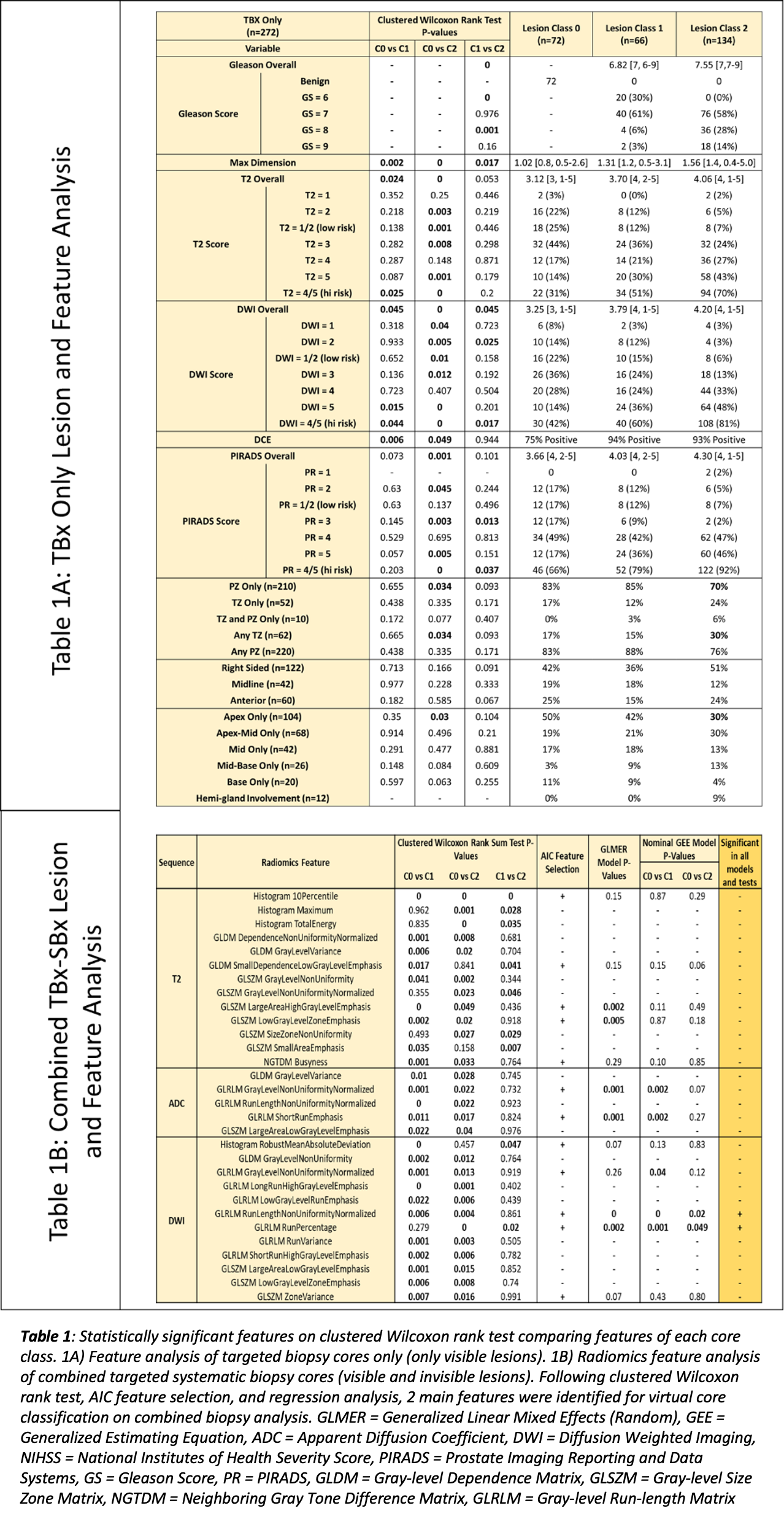
Introduction: Cribriform (CBFM) pattern on prostate biopsy has been implicated as a predictor for high-risk features, potentially leading to adverse outcomes after definitive treatment. Literature on imaging characteristics of CBFM is sparse and mixed. This study aims to elucidate the multiparametric MRI (mpMRI) features of CBFM-containing cancers on prostate biopsy using radiomics. Methods: Patients who underwent mpMRI, combined 12-core transrectal ultrasound (TRUS) guided systematic and MRI/US fusion-guided biopsy on a prostate cancer (PCa) clinical trial were retrospectively queried for the presence of CBFM pattern at biopsy. Biopsy cores were scored: C0=benign, C1=PCa with no CBFM pattern, C2=PCa with CBFM pattern. In addition to targeted lesion regions of interest (ROIs), patient-specific 12-core systematic TRUS biopsy sector maps were reconstructed and transposed on mpMRI slices for virtual core ROI creation (Figure 1). Radiomics features from each ROI were extracted using PyRadiomics package in Python. Radiomics and clinical feature analyses were done with Wilcoxon signed-rank test and Akaike Information Criterion. Results: Between 2020-2022, 90 consecutive patients with CBFM pattern on prostate biopsy and paired mpMRI were identified. Radiomics/clinical feature analysis included 1080 transrectal systematic biopsy cores (677 C0, 191 C1, 212 C2) and 272 MRI-targeted biopsy cores (72 C0, 66 C1, 134 C2). On combined biopsy analysis, two DWI radiomics features capturing signal heterogeneity were predictive for CBFM classification (p <.01). On analysis of MRI-targeted biopsy only, overall PIRADS score (p=.037), DWI PIRADS score (p=.017), and target lesion dimension (p=.017) were predictive for CBFM classification (Table 1). Conclusions: CBFM pattern has visible radiomics features on mpMRI, most notably on DWI. Imaging findings suggestive of CBFM may play a role in risk stratification and patient counseling. Further studies are needed to evaluate these lesions and validate their imageability longitudinally and externally. SOURCE OF Funding: None