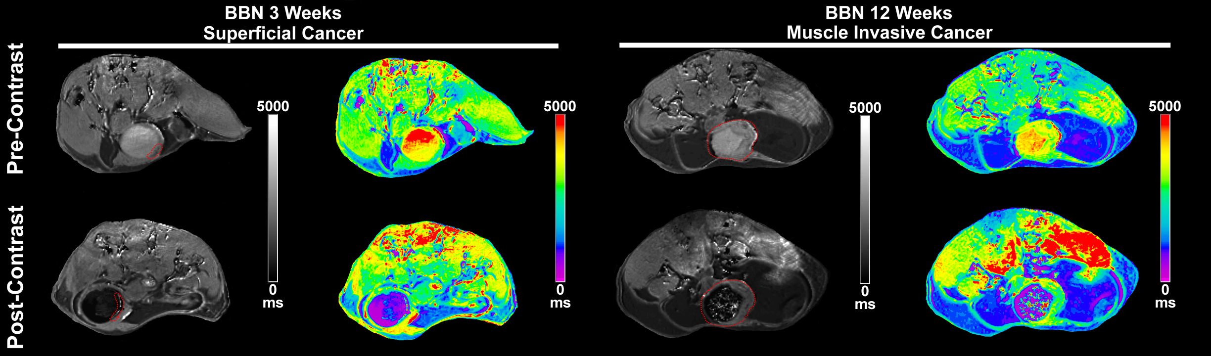
Introduction: Here, we examined the potential of intravesical contrast enhanced magnetic resonance imaging (ICE-MRI) for virtually monitoring the bladder tumor progression from 2-12 weeks, induced in mice fed water containing tobacco carcinogen, N-butyl-N-(4-hydroxybutyl) nitrosamine (BBN) that alkylates DNA to elicit the mutational burden and basal subtype cell markers of muscle invasive bladder cancer (MIBC) as well as reflect the four-fold higher cancer risk in past and current smokers. Methods: 6 weeks old immunocompetent female mice of B6D2F1 strain (n=20) were fed ad libitum water or water containing 0.05% BBN for upto 12 weeks and imaged by ICE-MRI at 7T under isoflurane anesthesia, before and after 0.05mL transurethral instillation of Gadobutrol (4mM) and Ferumoxytol (5mM) for a 30min period. Multi slice, axial T1 weighted images were acquired at variable repetition time of 400-7500ms for voxel-wise T1 mapping with echo time -6.5ms, slice thickness 0.8mm, field of view 28*28 mm2, acquisition matrix 218x218, and number of averages =1. Results: While urinary excretion of Gadobutrol injected i.v. for DCE-MRI generates pseudo layering in lumen, ICE-MRI accomplishes uniform image contrast in lumen with steep concentration gradient that resists dilution from fresh urine for size dependent paracellular diffusion of 0.8nm sized Gadobutrol while lumen is darkened from retention of 30nm Ferumoxytol. Paracellular diffusion exclusively of Gadobutrol and not Ferumoxytol across perturbed tight junction complex of cancerous cells shortens T1 relaxation times of water protons for differentiating normal bladder wall ( 2909 ms) from cancer foci (1048 ms) in color coded T1 maps (attached Fig). BBN fed mice exhibit superficial tumor after 2-3 weeks that progresses to MIBC stage (~4mm length) at multiple foci by 12 weeks in 50% of BBN mice having bladder wall twice the thickness of water fed mice. Conclusions: Here, we show that ICE-MRI can visualize small polyps and MIBC owing to tumoritropic infiltration of Gadobutrol. ICE-MRI leverages perturbed tight junction complex of cancer foci relative to normal regions for differential contrast enhancement to perform non-surgical surveillance of indolent cancer growth, detect recurrence and stage MIBC without transurethral resection. SOURCE OF Funding: NCI- CA263243, CA251341, P30CA047904