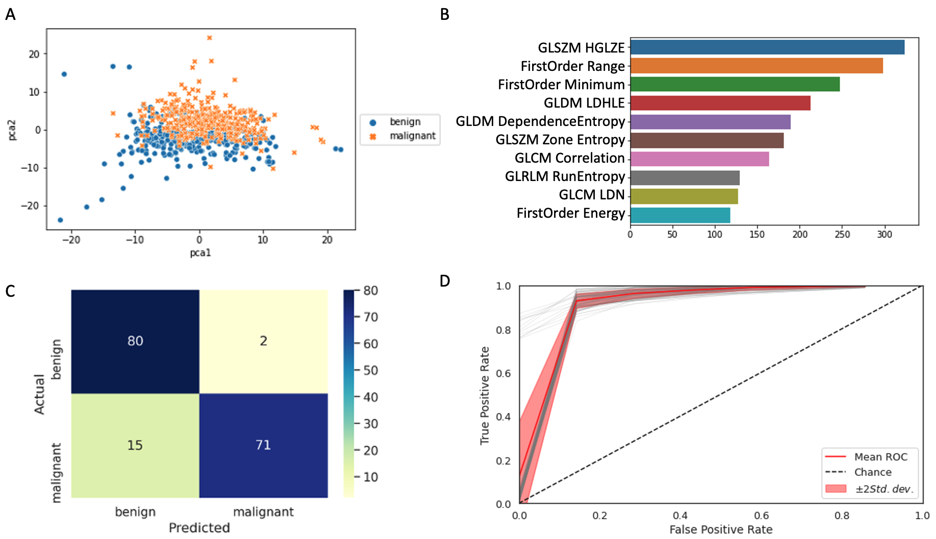
Introduction: Up to 20% of renal tumors are benign and may not require treatment. However, benign versus malignant renal tumors cannot be distinguished using cross-sectional imaging. Renal mass biopsy is a possible solution, but biopsy is invasive and has notable non-diagnostic and false negative rates. As a result, many patients proceed directly to treatment, including some who undergo extirpative surgery for benign tumors. We aim to develop a radiomics and machine learning model for distinguishing between oncocytoma versus malignant renal neoplasms based on abdominal CT images. Methods: Our institutional registry was queried for patients who underwent surgical treatment of renal tumors from 2000-2018. All surgical specimens underwent pathology re-review by an expert genitourinary pathologist. A total of 843 images from 609 patients (434 images of oncocytoma and 409 images of malignant renal tumor) were included. A previously developed artificial intelligence algorithm for segmentation of kidney, cyst, and tumor area was applied. Images were preprocessed by resampling via linear interpolation to 0.8 mm x 0.8 mm x 5 mm, window/level = 440/40, and intensity normalized [0 255]. Features were extracted for the tumor region using PyRadiomics with a fixed bin width of 16. Both unsupervised (PCA, tSNE) and supervised (LR, SVM) machine learning approaches were explored. Data was split 80:20, and cross validation was used in the training/validation set parameters. The best performing model based on F1 macro was then calibrated using an isotonic calibration approach. Results: Dimensionality reduction showed adequate class separation (Fig. 1A). The top 10 radiomic features showed a mixture of first and second order features (Fig. 1B). The final model reached an accuracy of 0.90 (Fig. 1C). Of the 168 images in the testing set, only 2 benign cases were predicted to be malignant, and 15 malignant cases were predicted to be benign. The AUC for oncocytoma versus malignant histology prediction was 0.96 with CI [0.93 0.98] (Fig. 1D). Conclusions: We developed a machine learning model for accurately distinguishing benign oncocytoma versus malignant renal tumors based on CT images alone. This may provide an opportunity for non-invasive risk stratification of solid renal neoplasms, which could potentially reduce overtreatment of renal tumors. SOURCE OF Funding: None

 photo")