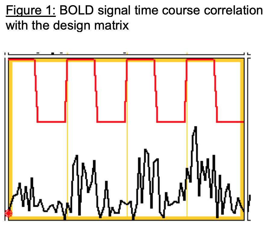
Introduction: Lower urinary tract (LUT) is under the control of the central nervous system. Functional MRI (fMRI) is designed to explore neuronal activity, however, its application at the level of the spinal cord has been challenging and not explored. The bulbocavernosus reflex (BCR) is a reliable means of assessing the integrity of the spinal cord, particularly at the sacral level of the LUT control. The BCR is usually triggered by a gentle squeezing of the penis glans or the clitoris, especially with a full bladder. A similar and more convenient method is to tap on the midline of the suprapubic region when the bladder is full. The objectives of this project are twofold: (1) to establish a feasible and MRI compatible BCR assessment within the MRI scanner, (2) to assess the feasibility of spinal cord fMRI during a full bladder BCR in regard to LUT control in healthy individuals. Methods: An MRI compatible hammer was designed and generated to deliver the stimulation within the MRI tunnel to elicit BCR. Two healthy males with no previous history of LUT symptoms or spinal cord injury were recruited for this trial. The participants were instructed to consume 500ml of water and empty their bladder immediately before entering the MRI. When they reported a full bladder, they underwent a task-based fMRI of four blocks of 40 seconds of tapping (BCR) and 40 seconds of control (rest). The regions of interest (ROIs) involved in the BCR, were defined as the area of the terminal cone (S1-S4 roots) known to be involved in LUT control. The blood level oxygen-dependent (BOLD) signal within ROIs was analyzed with the FSL software. Results: The ROIs identified in the sacral spinal cord showed a significant increase in signal upon BCR stimulation. BOLD activations were present and time-related to the stimulation period (Figure 1). Conclusions: This is the first study to demonstrate that: There is a feasible and modest approach to elicit BCR within the MRI to assess spinal cord integrity in regard to LUT control. The activity of the sacral spinal cord during the BCR stimulation can be revealed in fMRI. These preliminary results pave the way for further study of the functional role of the spinal cord in larger cohorts of healthy individuals and in individuals with spinal cord injury. SOURCE OF Funding: Funding provided by the National Institute of Health, NIDDK R03DK126994-01 award and the CAIRIBU Core collaboration award