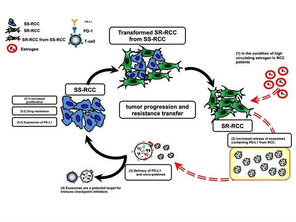
Introduction: In 2022, SEER data projects 79,000 new cases renal cell carcinoma (RCC) with 15% of these presenting with progressed disease and distant metastasis. Prior publications have indicated that estrogen plays an important role by increasing RCC risk in female obese patients (Sun et al, EBioMedicine, 2018). However, the role of estrogen is still unknown in the setting of metastatic disease and treatment resistance. The aim of this study was to investigate whether estrogen’s role in treatment resistance could be linked to exosomal transfer of this hormone. Methods: Overall survival data was extracted and analyzed from the TCGA based on several gene expression profiles. 786-O, 786-O Suni Resistant (SR), A498, A498 SR, Caki-2, Caki-2 SR and 293-T cells were cultured, exosomes collected, and later isolated using differential ultracentrifugation. After exposure to estrogen, downstream analysis included western blot and qNano exosome quantification. Statistical analysis was performed using R computational language. Results: TCGA database analysis revealed that upregulated ER-beta expression is associated with a 25% decrease in overall survival at 5 years. Increased ER-Beta was also linked to patients with increased stage and metastatic disease. After exposing our cell lines to estrogen for 24hr ER-beta, activated NF-kB, and PD-L1 expression increased in all 3 TKI-sensitive and resistant cell lines. However, when quantifying exosome concentrations after estrogen exposure our 3 TKI-sensitive lines displayed a decrease in exosome production. On the other hand, each of the TKI-resistant cells showed a 3-4 fold increased exosome concentrations. We then linked this discordance in exosome production with a similar pattern in EGFR expression. Conclusions: In conclusion, estrogen is an important molecule present in many patients with RCC. This study’s data shows that estrogen is a causative agent of increasing PD-L1 expression potentiated through exosome production. A graphical hypothesis based on this studies data of estrogen's role in the tumor microenvironment can be found in Figure 1. SOURCE OF Funding: U54 GM104940