Session: MP05: Stone Disease: Basic Research & Pathophysiology
MP05-20: Kidney stone growth ‘rings’ visualized using micro CT and fluorescence microscopy: Similarities in growth of Randall’s plaque calcium oxalate stones from the same kidneys
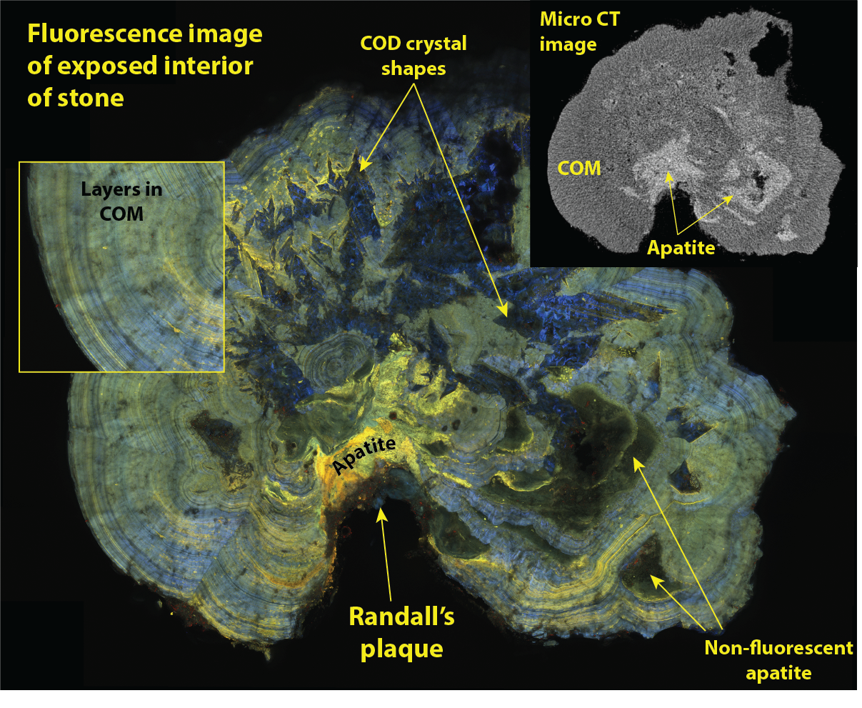
Introduction: Recent work has shown that mechanisms of stone growth can be demonstrated at the microscopic level using fluorescence microscopy. The objective here was to study multiple stones to observe patterns of mineral and organic deposition within and between patients. Methods: Stones were removed by basket during endoscopic procedure, and a total of 12 stones were studied from 3 patients. All stones were verified by micro CT as having grown on Randall’s plaque by residue visible on the stone. Each stone was mounted on polystyrene and ground down to reveal a planar surface inside the stone. The stone was then imaged using confocal microscopy (Leica SP8) with a water immersion lens (20x, 0.75 NA). Each stone was ground repeatedly to collect as many interior planes as possible, with micro CT verification of each plane of section and mineral regions exposed. Results: Within a patient, similarities of layering (both thickness and color) were apparent in stones from the same kidney, but stones from different patients had different fluorescent features. Several stones showed evidence of episodic growth by deposition of aggregates of calcium oxalate dihydrate crystals, and these stones with evidence of dihydrate aggregates tended to be larger stones within a given patient. Regions of apatite adjacent to Randall’s plaque showed yellow fluorescence distinctly different from apatite in later stone overgrowth. Conclusions: The unique fluorescence of apatite laid down as the first overgrowth on Randall’s plaque is suggestive of special urine molecules deposited at the initiation of stone growth. Outside of this consistent finding, fluorescent molecules laid down with mineral in calcium oxalate stones are not universal among patients, but stones from the same kidney showed similar fluorescence patterns. SOURCE OF Funding: NIH R01 DK124776, NIH P01 DK056788