Associate Professor Department of Nephro-urology, Nagoya City University Graduate School of Medical Sciences
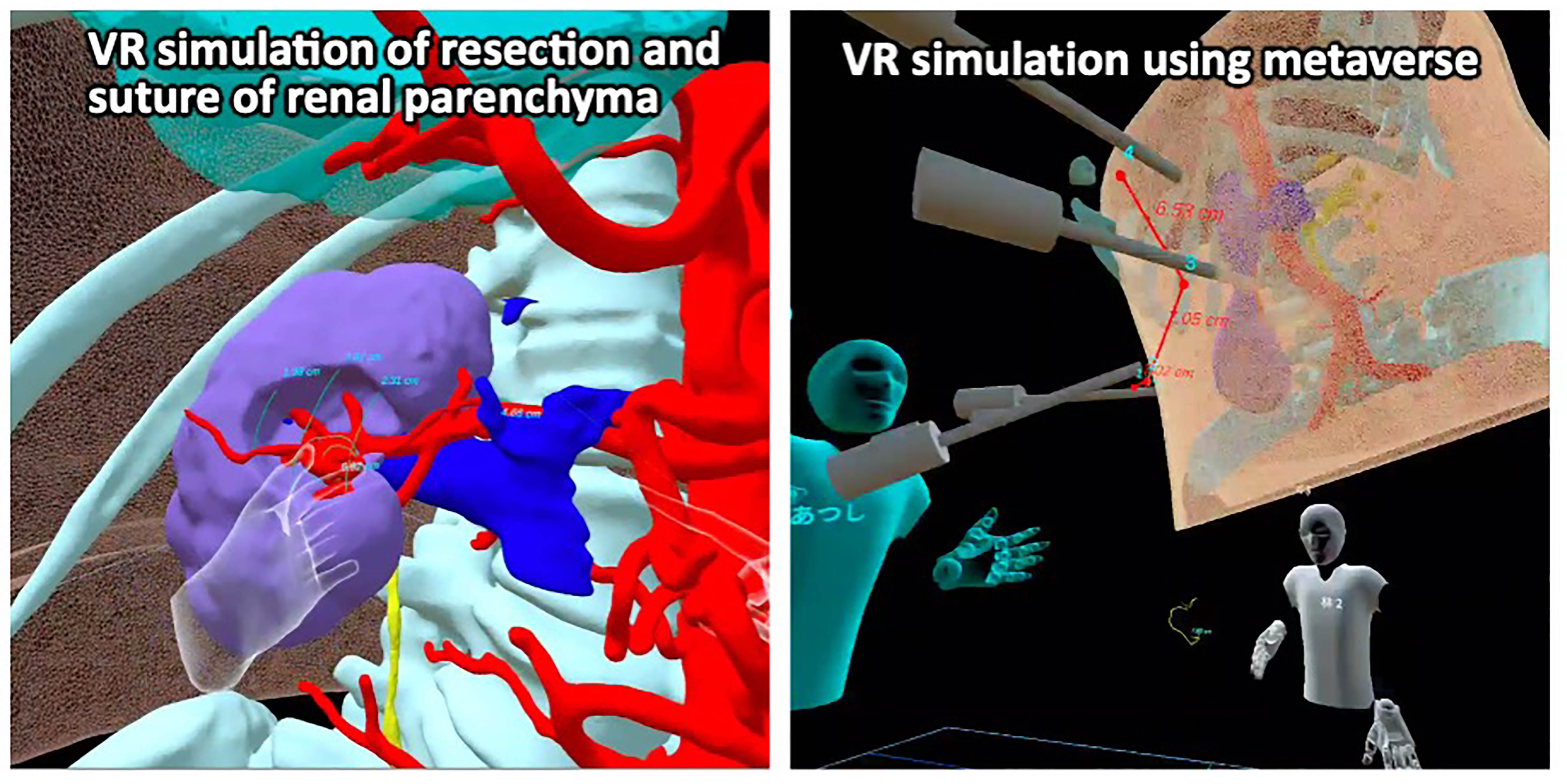
Introduction: We have developed a virtual reality (VR) simulation for safe performance for robot-assisted partial nephrectomy (RAPN). In order to share VR images with the surgical team, we developed a simulation that utilizes the virtual space (Metaverse). Methods: Stereolithography (STL) files of skin, bone, renal parenchyma after nephrectomy, tumor, resection margin, artery, vein, and urinary tract were created from preoperative contrast-enhanced CT using the Ziostation®2 medical imaging workstation. Along with these files, we also integrated the STLs of 4 virtual trocars and a 7 cm virtual sphere for port-to-port measurements in the application Holoeyes XR. Three to four doctors wore immersive 3D goggles and became avatars in the metaverse to observe patient anatomy constructed in VR. Results: In the Metaverse, not only did all doctors simultaneously observe the patient's unique three-dimensional anatomy as avatars, but there were also scenes where instructors instructed surgeons on approach methods. We were able to discuss the method of suturing from the location of the vessels and urinary collecting system exposed on the resected surface by partial nephrectomy. In addition, the virtual trocars could be used to determine the optimal port position for da Vinci operation. Conclusions: VR simulation for RAPN using Metaverse was shown to be useful not only for achieving safe surgery by sharing information about the surgical process with the team, but also for surgeon education. SOURCE OF Funding: Japanese Society of Endourology Grant

 photo")