MP73-13: MRI analysis of prostate morphometry and periprostatic anatomical relationships before and after partial high-intensity focused ultrasound treatment of localized prostate cancer.
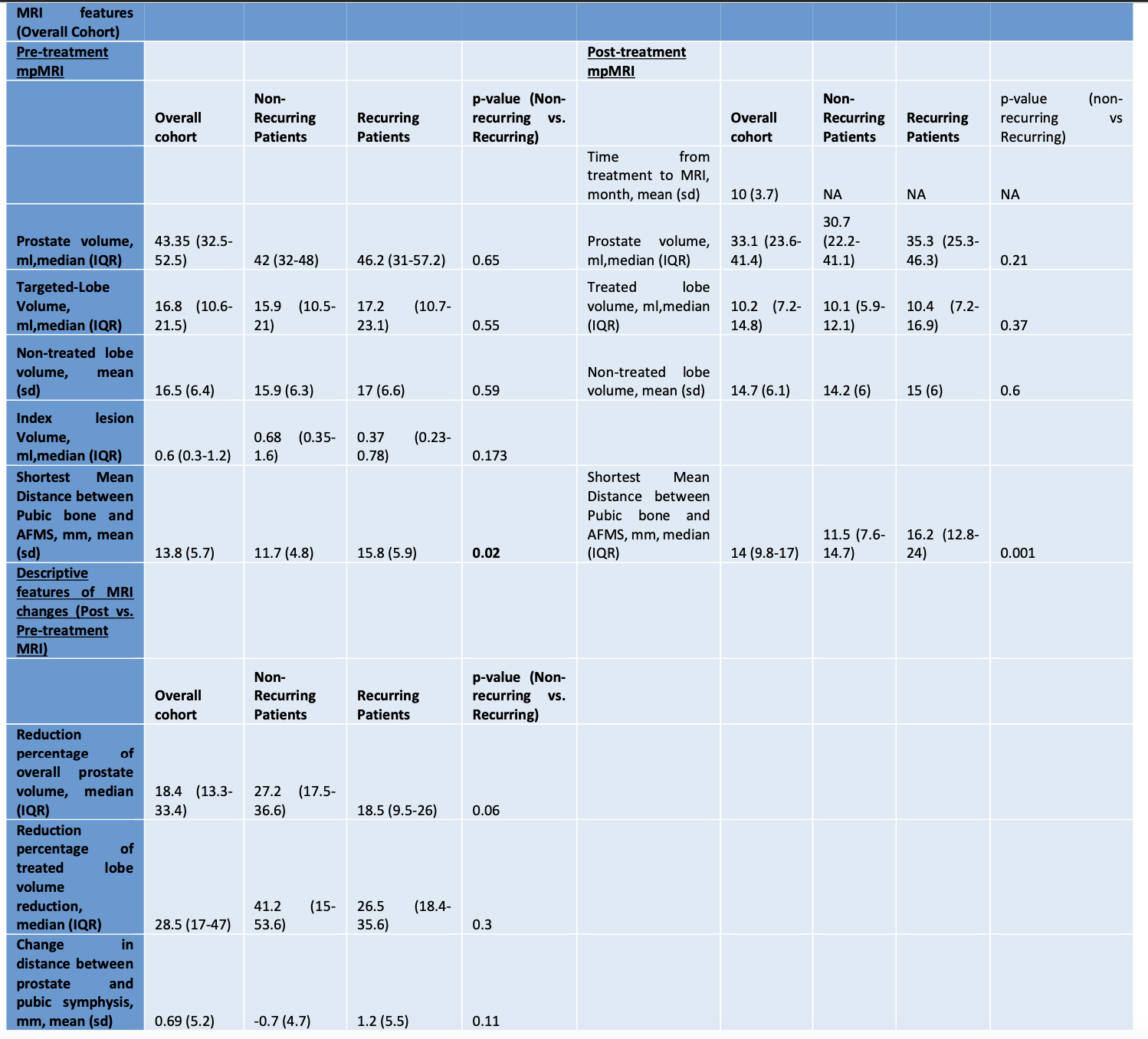
Introduction: Little is known regarding anatomical changes to peri-prostatic tissues after focal high intensity focused ultrasound (fHIFU) for localized prostate cancer. We sought to provide a novel approach to assess prostato-pelvic anatomical morphometric magnetic resonance imaging (MRI) changes post- fHIFU. Methods: 38 patients (Pts) over two institutions (Lille University Hospital and Duke University Hospital) undergoing fHIFU with pre-and post-treatment (Pre-/Post-Tx) (=6 months) MRI were included. Pre- and post-TX. MRIs were assessed for prostate and treated region volume. Distances to pubic bone and rectum were measured to account for the post-Tx periprostatic changes. Two subgroups were also identified, those with recurrence (positive biopsy and/or subsequent whole gland Tx) and those recurrence-free. Specific MRI features were also described within those two groups. Results: The median treated-lobe volume was of 16.8 ml (IQR 10.6-21.5) on pre-Tx MRI and of 10.2 ml on post-Tx MRI (IQR 7.2-14.8). Comparison on pre-Tx MRI vs. post-Tx MRI showed a significant difference (p < 0.01). The median percentage reduction of treated lobe volume for the entire cohort was 28.5% (IQR: 17-47). No significant difference was identified between the non-recurring group and the recurring group for prostate volume and treated lobe-volume both at pre- and post-Tx. Analysis of prostato-rectal measurements was conducted in 17 Pts. The mean value of the median prostato-rectal distance was 2 mm (SD 1) in pre-Tx and 1.97 mm (SD 1.1) in post-Tx. We noted no significant difference in either the pre-Tx or post-Tx configuration (p=0.78 and p=0.1 respectively) between the groups of recurring vs. non-recurring Pts. 13 Pts were qualitatively classified as presenting an aspect of loss of symmetry in the prostato-rectal space on post-Tx MRI. No significative difference existed between the recurring and non-recurring group for this parameter. Conclusions: With this study, we described a novel standardized method of morphometric alteration post-fHIFU. This preliminary work incite further studies to assess the clinical significance and implications of these results in a larger prospective cohort. SOURCE OF Funding: None