
.jpg)
Syndromes/Craniofacial Anomalies
Nicholas M. Carlson, DMD
Pediatric Dental Resident, PGY-2
Indiana University, Riley Hospital for Children
Indiana University, Riley Hospital for Children
Indianapolis, Indiana, United States
LaQuia A. Walker-Vinson, DDS, MPH, FAAPD
Associate Professor
Indiana University School of Dentistry / Riley Hospital for Children
Indianapolis, Indiana, United States
LaQuia A. Walker-Vinson, DDS, MPH, FAAPD
Associate Professor
Indiana University School of Dentistry / Riley Hospital for Children
Indianapolis, Indiana, United States
Treatment Management of a Pediatric Patient with Complex Oligodontia
Nicholas Carlson, DMD • LaQuia Vinson, DDS, MPH
Indiana University School of Dentistry • Riley Hospital for Children • Indianapolis, Indiana
Introduction:
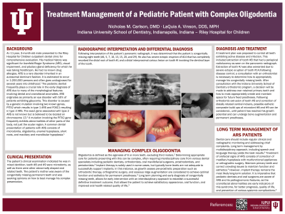
Axenfeld-Rieger Syndrome (ARS) is a rare disorder inherited in an autosomal dominant fashion. It is estimated to occur in 1:200,000 persons and often goes undiagnosed for several years into childhood. The pediatric dentist frequently plays a crucial role in the early diagnosis of ARS due to many of the morphological features involving dental and craniofacial anomalies. ARS originates as primarily an eye disorder with 50% of patients exhibiting glaucoma. This disorder is caused by a genetic mutation involving two known genes, PITX2 which results in type 1 ARS and FOXC1 resulting in type 3 ARS. The exact gene associated with type 2 ARS is not known but is believed to be located on chromosome 13. A mutation involving the PITX2 gene frequently exhibits abnormalities of other parts of the body, not just the ocular region. A common dental presentation of patients with ARS consists of microdontia, oligodontia, enamel hypoplasia, short roots, and maxillary and mandibular hypoplasia.
Case Study:
An 11-year, 3-month-old male presented to the Riley Hospital for Children outpatient dental clinic. His medical history was significant for Axenfeld-Rieger Syndrome (ARS), visual impairment, and pituitary gland deficiency for which, he was taking Norditropin. He had no known drug allergies. The patient presented with mixed dentition, teeth #8 and #9 were microdonts, as well as there were other abnormally shaped and rotated teeth. Following interpretation of the panoramic radiograph, it was determined that the patient was congenitally missing teeth #4, 5, 7, 10, 13, 15, 20, and 29. This report will review management of this patient with oligodontia associated with ARS as well as recommendations for patients with complex oligodontia.