Back

Clinical Research
(CR-027) Assessment of burn wounds using hyperspectral imaging

Co-Author(s):
Robert Bartlett, MD, CPE, MAPWCA, FAPWH, UHM, FACEP – Swift Medical; Sheila Wang, PhD, MD – McGill University and Swift Medical; Robert Fraser, MN, RN – Swift Medical; Amy Lorincz, BEng, MS – Swift Medical; Gennadi Saiko, PhD – Swift Medical; Mario Martinez-Jimenez, MD, PhD – Universidad Autonoma de San Luis Potosi
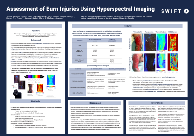
<b>Introduction</b>: <strong id="docs-internal-guid-e73bef06-7fff-a75a-6998-b426762c5e54" style="font-weight: normal;"><span style="font-size: 11pt; font-family: Roboto,sans-serif; color: #000000; background-color: transparent; font-weight: 300; font-style: normal; font-variant: normal; text-decoration: none; vertical-align: baseline; white-space: pre-wrap;">Hyperspectral imaging is the simultaneous acquisition of images at different wavelengths of the electromagnetic spectrum. This technique assesses perfusion, inflammation, and the infectious status of a wound. Burns are especially well suited for hyperspectral imaging, as their management requires confirmation of a clean wound with a high healing potential before surgical treatments can be done. Therefore, the objective of this study was to use a novel hyperspectral imaging device to assess burns before and after skin grafting and identify those image patterns that correlate with the need for surgical management and higher graft survival.</span></strong><br/><br/><b>Methods</b>: <strong id="docs-internal-guid-a780ac32-7fff-64f4-cfab-e1192d3b8eec" style="font-weight: normal;"><span style="font-size: 11pt; font-family: Roboto,sans-serif; color: #000000; background-color: transparent; font-weight: 300; font-style: normal; font-variant: normal; text-decoration: none; vertical-align: baseline; white-space: pre-wrap;">Burn victims were assessed immediately after admission into a burn unit and then every 3 to 5 days using a hyperspectral imaging device. Photographs with the wound area and tissue composition calculation, infrared thermal images, bacterial fluorescence, and oxyhemoglobin concentration image maps were obtained on each measurement. The patients' outcomes were recorded as either reepithelizing or requiring surgical management, and in the latter case, the percentage of graft survival was also recorded. Data was extracted from the different images obtained and compared across treatment groups.</span></strong><br/><br/><b>Results</b>: <strong id="docs-internal-guid-8b6fef6e-7fff-d474-8564-7d5341102034" style="font-weight: normal;"><span style="font-size: 11pt; font-family: Roboto,sans-serif; color: #000000; background-color: transparent; font-weight: 300; font-style: normal; font-variant: normal; text-decoration: none; vertical-align: baseline; white-space: pre-wrap;">Burns that reepithelized without surgical treatment and those with the highest graft uptake were richer in granulation tissue, hotter, and had higher oxyhemoglobin concentrations. In all cases, confirmation of the absence of bacterial growth was done using the hyperspectral imaging device.</span></strong><br/><br/><b>Discussion</b>: <p dir="ltr" style="line-height: 1.38; margin-top: 0pt; margin-bottom: 0pt;"><span style="font-size: 11pt; font-family: Roboto,sans-serif; color: #000000; background-color: transparent; font-weight: 300; font-style: normal; font-variant: normal; text-decoration: none; vertical-align: baseline; white-space: pre-wrap;">Hyperspectral imaging offers a deep insight into the burn healing process and allows the identification of patients with higher success for reepithelization or skin grafting uptake. This technology is a non-contact, non-invasive, point-of-care imaging device that allows for monitoring of the healing response and the inflammatory or infectious status of burns to better support clinical decision-making. </span></p><br/><br/><b>Trademarked Items</b>: <br/><br/><b>References</b>: Ramirez-GarciaLuna JL, Bartlett R, Arriaga-Caballero JE, Fraser RDJ, Saiko G. Infrared Thermography in Wound Care, Surgery, and Sports Medicine: A Review. Frontiers in Physiology (2022) [cited 2022 Mar 12];13. Available from: https://www.frontiersin.org/article/10.3389/fphys.2022.838528
Oropallo AR, Andersen C, Abdo R, Hurlow J, Kelso M, Melin M, et al. Guidelines for Point-of-Care Fluorescence Imaging for Detection of Wound Bacterial Burden Based on Delphi Consensus. Diagnostics (Basel). 2021 Jul 6;11(7):1219.
Saiko, G., Lombardi, P., Au, Y., Queen, D., Armstrong, D., and Harding, K. (2020). Hyperspectral imaging in wound care: A systematic review. International Wound Journal 17, 1840–1856. doi: 10.1111/iwj.13474.<br/><br/>

.png)