Back

Clinical Research
(CR-028) Is my wound infected? Use of hyperspectral imaging to assess a wound’s infectious status

Co-Author(s):
Mario Martinez-Jimenez, MD, PhD; Robert Bartlett, MD, CPE, MAPWCA, FAPWH, UHM, FACEP – Swift Medical; Sheila Wang, MD, PhD – McGill University & Swift Medical; Robert Fraser, MN, RN – Swift Medical; Amy Lorincz, BEng, MS – Swift Medical; Gennadi Saiko, PhD – Swift Medical; Gregory Berry, MDCM, MSEd, FRCSC – McGill University
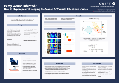
<b>Introduction</b>: <strong id="docs-internal-guid-0b341796-7fff-0957-4b61-efba75ac614c" style="font-weight: normal;"><span style="font-size: 11pt; font-family: Roboto,sans-serif; color: #000000; background-color: transparent; font-weight: 300; font-style: normal; font-variant: normal; text-decoration: none; vertical-align: baseline; white-space: pre-wrap;">Wound infections occur in a spectrum that goes from contamination, colonization, local infection, spreading infection, to systemic infection. A common challenge is discriminating between those wounds contaminated and colonized from those with subtle local infections. Hyperspectral wound imaging can help identify inflammation and bacterial presence on wounds, thus helping categorize them as being infected, inflamed or non-inflamed.<br /><br /></span></strong><strong id="docs-internal-guid-0b341796-7fff-0957-4b61-efba75ac614c" style="font-weight: normal;"><span style="font-size: 11pt; font-family: Roboto,sans-serif; color: #000000; background-color: transparent; font-weight: 300; font-style: normal; font-variant: normal; text-decoration: none; vertical-align: baseline; white-space: pre-wrap;"><br /></span></strong><br/><br/><b>Methods</b>: <strong id="docs-internal-guid-b8667431-7fff-3f0e-8022-19d39697ae0c" style="font-weight: normal;"><span style="font-size: 11pt; font-family: Roboto,sans-serif; color: #000000; background-color: transparent; font-weight: 300; font-style: normal; font-variant: normal; text-decoration: none; vertical-align: baseline; white-space: pre-wrap;">A series of 64 patients with wounds suspicious of infections were imaged using a novel hyperspectral imaging device. An infectious process was confirmed either by tissue biopsy microbiological culture or clinically on-site by an expert surgeon. Analysis of the images was performed and the features that correlated with an inflammatory verses infectious process are presented here. <br /><br /><br /><br /></span></strong><br/><br/><b>Results</b>: <p dir="ltr" style="line-height: 1.38; margin-top: 0pt; margin-bottom: 0pt;"><strong id="docs-internal-guid-76028b1e-7fff-7596-fd9f-32e3bdb0813f" style="font-weight: normal;"><span style="font-size: 11pt; font-family: Roboto,sans-serif; color: #000000; background-color: transparent; font-weight: 300; font-style: normal; font-variant: normal; text-decoration: none; vertical-align: baseline; white-space: pre-wrap;">Qualitative analysis of the images showed four patterns, with two of them considered clinically or microbiologically as infected. One pattern consisted of wounds with a slightly increased temperature in wound bed area, increased peri-wound temperature, and negative to slightly positive bacterial fluorescence. The other pattern included wounds with a slightly increased wound bed temperature, increased peri-wound temperature, and positive bacterial fluorescence. Two non-infected patterns were seen, though have not been included in the abstract due to word count considerations.<br /><br /></span></strong><strong id="docs-internal-guid-76028b1e-7fff-7596-fd9f-32e3bdb0813f" style="font-weight: normal;"><span style="font-size: 11pt; font-family: Roboto,sans-serif; color: #000000; background-color: transparent; font-weight: 300; font-style: normal; font-variant: normal; text-decoration: none; vertical-align: baseline; white-space: pre-wrap;"><br /></span></strong></p><br/><br/><b>Discussion</b>: <strong id="docs-internal-guid-665f4c81-7fff-ca3c-eb46-75481449dd86" style="font-weight: normal;"><span style="font-size: 11pt; font-family: Roboto,sans-serif; color: #000000; background-color: transparent; font-weight: 300; font-style: normal; font-variant: normal; text-decoration: none; vertical-align: baseline; white-space: pre-wrap;">The combined use of clinical data plus hyperspectral images, including thermal and bacterial fluorescence imaging, can be used to discriminate between aseptic inflammation and wound infection. Hyperspectral imaging can be used to complement a patient’s clinical assessments and enhance clinical decision making.<br /><br /><br /><br /></span></strong><br/><br/><b>Trademarked Items</b>: <br/><br/><b>References</b>: Gentry EM, Kester S, Fischer K, Davidson LE, Passaretti CL. Bugs and Drugs: Collaboration Between Infection Prevention and Antibiotic Stewardship. Infect Dis Clin North Am. 2020 Mar;34(1):17–30.
Ramirez-GarciaLuna JL, Bartlett R, Arriaga-Caballero JE, Fraser RDJ, Saiko G. Infrared Thermography in Wound Care, Surgery, and Sports Medicine: A Review. Frontiers in Physiology (2022) [cited 2022 Mar 12];13. Available from: https://www.frontiersin.org/article/10.3389/fphys.2022.838528
Oropallo AR, Andersen C, Abdo R, Hurlow J, Kelso M, Melin M, et al. Guidelines for Point-of-Care Fluorescence Imaging for Detection of Wound Bacterial Burden Based on Delphi Consensus. Diagnostics (Basel). 2021 Jul 6;11(7):1219.<br/><br/>

.png)