Back
Discovery and Basic Research
Session: Symposium: Oncotargets: Challenges & Opportunities
High-Throughput 3D Bio-Printed Tumor Microenvironment Platform for Mimicking Lung Cancer
Monday, October 17, 2022
10:30 AM – 11:00 AM ET
Location: 255

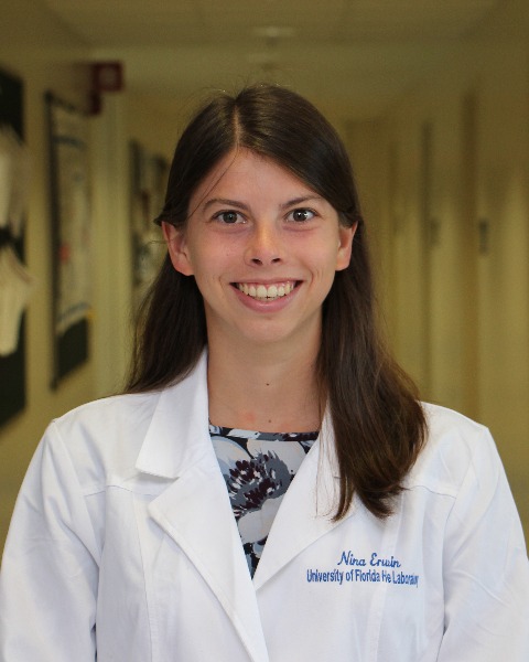
Nina Erwin
Graduate Student
University of Florida
Gainesville, Florida
Speaker(s)
Tumor microenvironments contribute to the complex molecular and phenotypic cellular changes within a solid tumor. Thus, more robust in vitro tumor microenvironment models are critical for cancer research and identifying clinically effective drug candidates. 3D multicellular tumor spheroids can more closely mimic the in vivo vascularized tumors and are gaining popularity for high-throughput screening and high-yield success for anti-cancer drug development. However, the conventional methods for tumor spheroid generation are laborious and low throughput and result in the inconsistent quality of spheroids, leading to unreliable tumor tissue models. Therefore, more attention is being given to the fabrication of in vitro 3D tumor models to study the model-dependent in-vitro cytotoxicity assay development and cancer immunotherapy. Ongoing studies integrate tumor microvasculature and tumor–stromal microenvironment with high throughput screening capability via microfluidic tumor-collagen matrix droplet generation for the 3D culture of tumor cells to mimic the tumor microenvironment. In this research, we have demonstrated that the high throughput, large-scale 3D collagen matrix spheroids can be used as the bio-ink for extrusion-based 3D bioprinting and build high shape fidelity and in vivo like 3D lung tumor models. Thus, harnessing the power from both 3D bioprinting and multicellular spheroids via a microfluidic platform can lead to the development of highly effective and reliable tumor models, opening up another road in the precise understanding of the mechanism of tumor progression and revolutionizing anti-cancer drug discovery process.
Learning Objectives:
- Upon completion, the participant will be able to obtain knowledge on the microfluidic generation of 3D tumor spheroids to mimic the tumor microenvironment for biomedical applications
- Upon completion, the participant will be able to gather sufficient information on using 3D bioprinting as a novel tool to develop tumor microenvironment and biomimetic tissue for long-term study
- Upon completion, the participant will be able to compare the biomimetic tissue to in vivo tissue environment

