Back
Poster, Podium & Video Sessions
Moderated Poster
MP32: Surgical Technology & Simulation: Instrumentation & Technology I
MP32-14: In Vitro Testing of Untethered Thermal-Actuated Microgrippers as a Novel Diagnostic Tool for Urothelial Tissue
Saturday, May 14, 2022
2:45 PM – 4:00 PM
Location: Room 225
Ridwan Alam*, Wangqu Liu, Ezra Baraban, Brian Matlaga, Andres Matoso, David Gracias, Baltimore, MD, Jared Winoker, New York City, NY

Ridwan I. Alam, MD,MPH
Johns Hopkins University School of Medicine
Poster Presenter(s)
Introduction: The gold standard for diagnosis of a suspicious upper tract mass is ureteroscopic biopsy. However, due to the microscopic caliber of the ureter, the development of tools to obtain an adequate tissue sample for analysis has proven challenging. All tools to date have required tethering to the ureteroscope, which severely restrict the size of the biopsy instrument and range of motion. As a result, up to 65% of upper tract malignancies are inaccurately staged. Therefore, we propose the development of untethered submillimeter tools called microgrippers to obtain tissue samples from the upper urinary tract using an in vitro model of porcine urothelium.
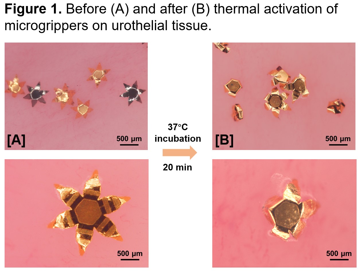
Methods: Microgrippers are microsurgical tools measuring 1 mm in the open configuration (Figure 1A). These devices are fabricated using materials safe for use within the human body. A thermosensitive layer keeps the microgrippers in the open configuration at 4°C but softens at 37°C, allowing the microgrippers to close (Figure 1B). The closing motion allows the device to capture contents within its phalanges. Experiments were conducted by placing microgrippers on a sample of porcine urothelial tissue. The microgrippers were retrieved using a magnet and centrifuged to separate the microgrippers from tissue. Tissue samples were analyzed using standard histopathological techniques.
Results: Urothelial tissue was successfully obtained when tested on 10 separate samples of porcine ureteral and renal pelvic tissue. Histological analysis confirmed the presence of urothelial tissue without excessive damage or artifact.
Conclusions: Untethered thermal-actuated microgrippers demonstrate the ability to adequately sample urothelial tissue in vitro for analysis by pathologists. There is potential for this revolutionary technology to establish a new standard for biopsy of the upper urinary tract. Ongoing studies are focused on ex vivo and in vivo experimentation for biopsy and drug delivery mechanisms.
Source of Funding: None.


.jpg)
.jpg)