Back
Poster, Podium & Video Sessions
Moderated Poster
MP36: Renal Transplantation & Vascular Surgery
MP36-20: Multi-Photon Microscopy for the Evaluation of Interstitial Fibrosis in Extended Criteria Donor Kidneys: A Proof-of-Concept Pilot Study
Sunday, May 15, 2022
7:00 AM – 8:15 AM
Location: Room 228
Wei Zheng So, Jirong Lu*, Benjamin Yen Seow Goh, Rachel Zui Chih Teo, Anantharaman Vathsala, Ho Yee Tiong, Kent Ridge, Singapore
- JL
Poster Presenter(s)
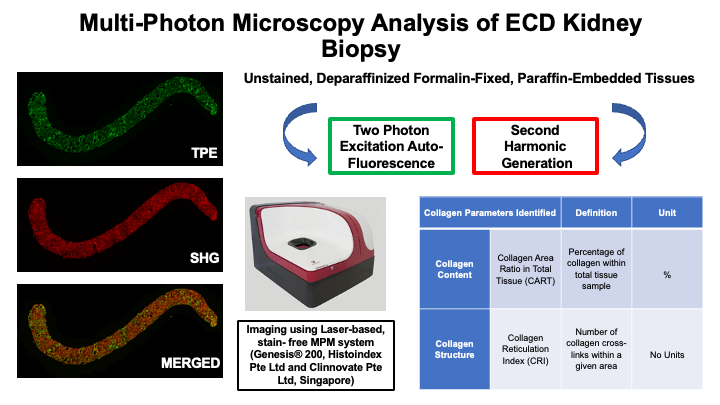
Introduction: This study evaluated the initial use of label free second harmonic generation (SHG) imaging with two photon excitation (2PE) auto-fluorescence for the quantification of collagen/fibrosis on pre-implantation biopsies of expanded criteria donors (ECD).
Methods: A total of 22 core ECD kidney transplant biopsy specimen tissues from 7 ECD donors, taken at the time of pre-implantation were retrieved, cut into 5-micron sections, mounted on slides and deparaffinized. The core needle biopsies were imaged with 2X and 20X objective using the commercially available laser-based Genesis® 200 (Histoindex Pte Ltd and Clinnovate Pte Ltd, Singapore). The entire core was selected as the Region of Interest (ROI). Corresponding clinical information from transplant donors were retrieved. Pathological review was performed, and all biopsies had Interstitial Fibrosis (IF)/Tubular Atrophy (TA) scores > 0. Collagen parameters measured included quantification by the Collagen Area Ratio in Tissue (CART), and qualitative measurements by Collagen Reticulation Index (CRI).
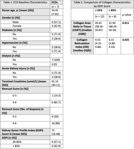
Results: 22 core biopsies were extracted from 14 donor kidney samples, of which originated from 7 donors. Table 1 depicts the baseline ECD characteristics of the donors. For the 8 kidneys with multiple biopsies done, we obtained an average score of the collagen parameters across the different samples. Biopsies classified with > 85% KDPI score had significantly higher CAR and CART than biopsies with = 85% KDPI score (Table 2).
Conclusions: MPM is an evolving technology that enables the quantification of the amount (CART) and quality (CRI) of collagen deposition in unstained explant biopsies of ECD kidneys. This initial evaluation found significant difference in both parameters between ECD donors with more or less than 85% KDPI.
Source of Funding: None


.jpg)
.jpg)