Back
Poster, Podium & Video Sessions
Moderated Poster
MP44: Stone Disease: Surgical Therapy (including ESWL) III
MP44-01: What is the ideal follow up after kidney stone treatment? A systematic review and follow-up algorithm from the European Association of Urology urolithiasis panel
Sunday, May 15, 2022
1:00 PM – 2:15 PM
Location: Room 228
Riccardo Lombardo*, Rome, Italy, Lazaros Tzelves, Athens, Greece, Robert Geraghty, Newcastle-upon-Tyne, United Kingdom, Niall F Davis, Dublin, Ireland, Andreas Neisius, Trier, Germany, Ales Petřík, Prague, Czech Republic, Giovanni Gambaro, Verona, Italy, Christian Türk, Vienna, Austria, Bhaskar Somani, Southampton, United Kingdom, Andreas Skolarikos, Athens, Greece, Kay Thomas, London, United Kingdom
- RL
Poster Presenter(s)
Introduction: Stone disease is a common and costly clinical condition, with currently no specific recommendations regarding when and how to follow-up these patients. The aim of our study was to develop a follow-up flow-chart after stone disease treatment, based on a systematic review performed by EAU Urolithiasis Guidelines Panel on urolithiasis follow-up.
Methods: The panel critically evaluated the raw data (stone free status, residual growth and spontaneous expulsion rate at different time points) and the outcome derived from a systematic review (PROSPERO: CRD42020205739) on urinary stone patients’ follow-up after definitive treatment. Given the lack of comparative studies between different follow-up modalities, a consensus statement between Panel members was made to establish the follow up of different group of patients. The final algorithm was developed using a benefit/harm principle. Every proposal of follow up was thoroughly discussed by all Panel members to finalize the algorithm.
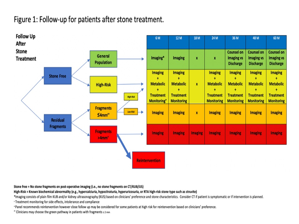
Results: A total of 76 studies were included in the analysis. In the stone free general population group, 71.4-100% of patients are stone-free at 12-months and 28.6-94% remain stone-free at 36 months. The stone-free rate in high-risk patients not under targeted medical therapy is <40% at 36 months. Patients with residual fragments =4 mm have a spontaneous expulsion rate of 17.9-46.5% and a growth rate of 10.1-40.7% at 12-months. Patients with residual fragments >4 mm should be considered for surgical re-intervention based on the high risk of recurrence (13-50% at 1 year). Risk of bias was moderate. A follow-up algorithm pertaining to the 4 sub-categories assessed is summarized in Figure 1. To develop the diagram all panel members agreed on following patients with plain film KUB and/or KUS based on clinicians’ preference and stone characteristics. However, NCCT scan should be performed if patient is symptomatic or if intervention is planned.
Conclusions: Based on the best evidence available we propose, for the first time, a possible follow-up chart for patients after stone surgical treatment. Despite existing limitations, this flow chart can improve the management of patients after stone disease treatment.
Source of Funding: None


.jpg)
.jpg)