Back
Poster, Podium & Video Sessions
Podium
PD31: Trauma/Reconstruction/Diversion: Urethral Reconstruction (including Stricture, Diverticulum) II
PD31-06: Expression and Localization of miR-146a-5p and miR-142-3p in Urethral Submucosa of Patients with Lichen Sclerosus-Induced Urethral Stricture Disease
Saturday, May 14, 2022
4:20 PM – 4:30 PM
Location: Room 252
Aaron Perecman*, Harjivan Kohli, Brandon Childs, Travis Sullivan, Burlington, MA, Myrtha Constant, Eric Burks, Boston, MA, Kimberly Rieger-Christ, Alex Vanni, Burlington, MA
- AP
Aaron Jacob Perecman, MD
Resident
Podium Presenter(s)
Introduction: In a prior study, we identified microRNA (miRNA) characteristic of lichen sclerosus (LS) induced urethral strictures (US). For this study, we used in situ-hybridization (ISH) to further investigate miRNA expression levels and localization in tissue from patients with LS-induced and non-LS US. ISH is a technique that detects and localizes DNA or RNA sequences in tissue by using labeled complementary probes. The goal of this study was to assess whether expression and localization of specific miRNAs were different between LS-induced and non-LS US.
Methods: 10 LS-induced and 9 non-LS US tissue samples were selected for ISH based on pathological and clinical review. 8 unique miRNAs identified from our prior study were chosen for ISH analysis. Tissue microarrays were prepared using 2mm punches of the stricture area. 5µm sections were stained using heat induced epitope retrieval with miRNAscope HD reagents (ACDBio, Newark, CA). Semi-quantitative tissue staining was scored by a single genitourinary pathologist for epithelial and submucosal regions (0= no detection, 1= low, 2= moderate, and 3= high expression). Data analysis was performed using SPSS v. 27. Variables were compared by the Wilcoxon rank sum test with significance considered at p<0.05.
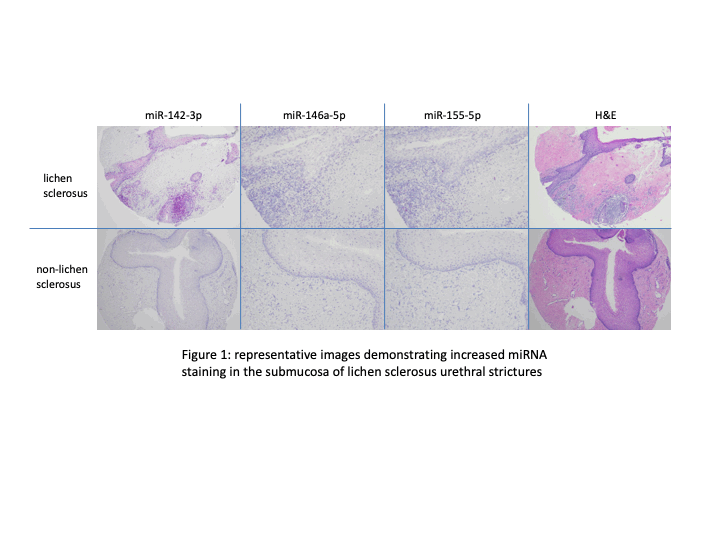
Results: There was significant difference in expression of miRNAs 146a-5p (p=0.028) and 142-3p (p=0.022) in the submucosa of LS-induced versus non-LS US. Representative images demonstrating increased staining for these miRNA probes are shown in Figure 1. MiRNA 155-5p likely had higher expression in the submucosa of LS tissue, but these results were not statistically significant (p < 0.08). There was no significant difference in miRNA expression in the epithelia of these samples.
Conclusions: Using ISH, this study showed up-regulation of several miRNAs in the submucosa of LS-induced US. The genetics of urethral LS remain under-investigated, and so establishing a miRNA profile may allow for better characterization of LS in order to ultimately guide prognosis and treatment. Further, histologic evaluation of LS is often difficult and these markers may aid in pathologic diagnosis.
Source of Funding: Robert E. Wise Research and Education Institute, Ellison Foundation.


.jpg)
.jpg)