Back
Poster, Podium & Video Sessions
Podium
PD40: Uroradiology II
PD40-04: How Does Magnetic Field Strength in Multiparametric Magnetic Resonance Imaging Affect Significant Prostate Cancer Detection? : A Multicenter Propensity Score–Matched Analysis
Sunday, May 15, 2022
10:00 AM – 10:10 AM
Location: Room 252
SUNAO SHOJI*, KUMPEI TAKAHASHI, SOICHIRO YUZURIHA, TATSUO KANO, IZUMI HANADA, TAKAHIRO OGAWA, TATSUYA UMEMOTO, MAYURA NAKANO, MASAYOSHI KAWAKAMI, MASAHIRO NITTA, MASANORI HASEGAWA, AKIRA MIYAJIMA, Isehara, Japan

Sunao Shoji, MD, PHD, MBA
Tokai University School of Medicine
Podium Presenter(s)
Introduction: This study aimed to analyze the effect of magnetic field strength of multiparametric magnetic resonance imaging (mpMRI) for the detection of clinically significant prostate cancer (SC) on MRI–transrectal ultrasound (TRUS) fusion image-guided target biopsy.
Methods: Patients whose serum prostate specific antigen (PSA) levels were = 20 ng/mL and who underwent MRI-TRUS fusion image-guided target biopsy in 2013–2021 were included. After propensity score matching was used to correct differences in patient age, PSA value, prostate volume, PSA density, highest PI-RADS category, and location of the cancer suspicious lesions with the highest PI-RADS category (transition zone [TZ] vs. peripheral zone [PZ]), the detection rates of SC among suspicious lesions with the highest PI-RADS category according to magnetic field strength of mpMRI (1.5 Tesla [T] vs. 3.0 Tesla [T]) were compared.
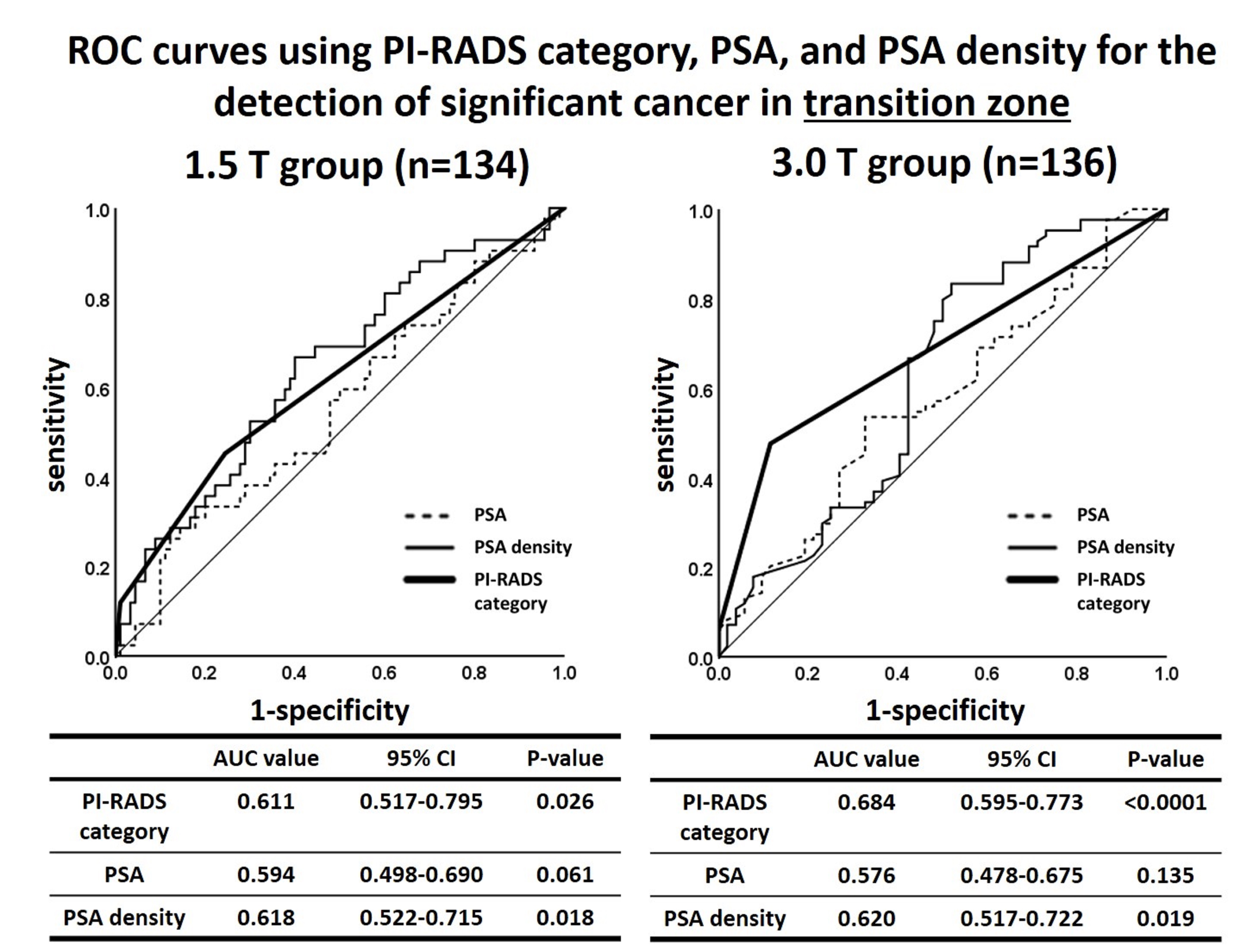
Results: Propensity score matching resulted in 257 patients in the 1.5 T group and 257 patients in the 3.0 T group. Median age, serum PSA level, and prostate volume of the 1.5 T and 3.0 T groups were 69 and 70 years (P=0.614), 8.07 and 8.06 ng/mL (P=0.), 30 and 30 cc (P=0.743), respectively. The detection rates of SC in the TZ in the 1.5 T and 3.0 T groups were PI-RADS category 3, 25.0% and 48.9% (P=0.001); PI-RADS category 4, 40.0% and 85.3% (P <0.0001); and PI-RADS category 5, 85.7% and 100% (P=0.755), respectively. The detection rates of SC in the PZ in the 1.5 T and 3.0 T groups were PI-RADS category 3, 63.2% and 70.9% (P=0.371); PI-RADS category 4, 93.2% and 94.9% (P=0.712); and PI-RADS category 5, 100% and 100% (P=0.712), respectively. Although the areas under the receiver operating characteristic curves (AUC) for the highest PI-RADS category were significantly greater than non-discrimination for the detection of SC in the TZ in the 1.5 T (AUC, 0.611; 95% CI, 0.517–0.705; P=0.026) and 3.0 T (AUC, 0.684; 95% CI, 0.595–0.773; P<0.0001) groups, the AUC was larger in the 3.0 T group than in the 1.5 T group.
Conclusions: A 3.0 T magnetic field on MRI yielded significantly higher detection rates of SC among suspicious cancer lesions classified as PI-RADS category 3 or 4 in the TZ.
Source of Funding: None.


.jpg)
.jpg)