Back
Poster, Podium & Video Sessions
Podium
PD47: Sexual Function/Dysfunction: Evaluation II
PD47-05: Serum Predictors of Pituitary Tumor Presence on MRI
Sunday, May 15, 2022
1:40 PM – 1:50 PM
Location: Room 245
Jose M Flores*, Nicole Benfante, John P Mulhall, NYC, NY
- JF
Podium Presenter(s)
Introduction: In clinical andrological practice, it is recommended that men with very low total testosterone (TT) levels as well as men with low TT/low LH with hyperprolactinemia should have a pituitary MRI to evaluate for a pituitary tumor (PT). What TT or prolactin levels mandate this, remains unclear. We aimed to define which TT and prolactin levels predicted PT presence on MRI.
Methods: The study population included men evaluated due to (i) low T (= 300 ng/dL) (ii) having early morning TT (iii) prolactin levels and (iv) brain/pituitary MRI. History of testosterone therapy or deprivation were exclusions. Early morning TT was measured using LCMS, and prolactin was assayed using an immunoassay. Prolactin level =20ng/mL was deemed high and TT = 150 ng/dL profoundly low T. The MRI was read by a neuroradiologist. We attempted to evaluate predictors of PT presence using multivariable analysis.
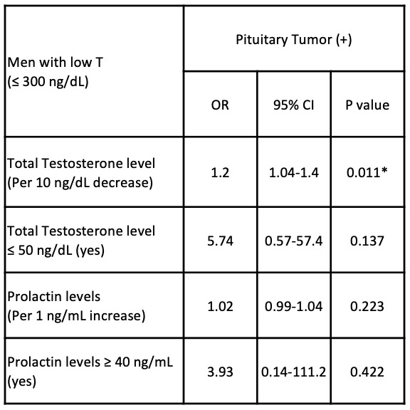
Results: 57 patients were included. Mean age was 56 ± 16 years. Mean TT = 146 ± 101 ng/dL and mean prolactin = 11 ± 11 ng/mL. A PT was diagnosed on MRI in 21% of these men. Median size was 0.7 (0.6, 1.4) cm. 58% had microadenoma, 33% macroadenoma and 9% other. 51% had profoundly low T and 16% high prolactin levels. 21% with TT < 300 ng/dL had a PT diagnosed; 38% profoundly low T; 30% in men with elevated prolactin. Lower TT levels (OR = 1.2) were the only predictor of PT.
Conclusions: In men with low T, 1 in 5 had PT, which increased to 1 in 3 in men with high prolactin levels and up to 40% in profoundly low T. We observed that only lower T increased the likelihood of PT.
Source of Funding: None


.jpg)
.jpg)