Back
Poster, Podium & Video Sessions
Moderated Poster
MP07: Bladder & Urethra: Anatomy, Physiology & Pharmacology
MP07-04: Development of a Porcine Model to Quantify Afferent Nerve Signaling: A Preliminary Study
Friday, May 13, 2022
10:30 AM – 11:45 AM
Location: Room 228
Christopher Bednarz*, Michael Shields, Graham Pingree, Abraham Alattar, David Lester, Ashley Cardenas, Sean Bryant, William Visser, John Speich, Adam Klausner, Richmond, VA
- CB
Christopher Paul Bednarz, MD
Virginia Commonwealth University Health System
Poster Presenter(s)
Introduction: Afferent nerve signals in response to bladder filling are a critical component in the normal micturition cycle. It is proposed that aberrations in signaling may be associated with voiding dysfunction. The purpose of this study was to develop a model to capture afferent nerve signals in perfused porcine bladders. To our knowledge this is a model that has not been previously reported.
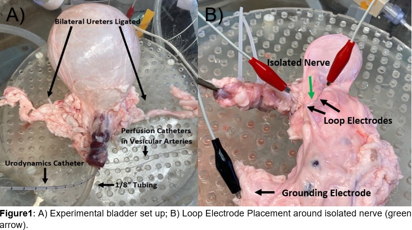
Methods: Porcine bladders were harvested fresh from local abattoirs. Ex vivo bladders were stored in physiologic buffer solution and prepared for experimentation by intubation of vesicular arteries, ligation of bilateral ureters, and cannulation of the urethra with both a Urodynamics catheter and a length of 1/8 inch tubing (Figure 1A). Nerves were isolated and loop electrodes were placed to monitor signaling activity using an AD Instruments PL2604 Power Lab (Figure 1B). To preserve bladder integrity, vesicular arteries were perfused with physiologic buffer solution. Bladders were filled with deionized water from a volume of 0 to 1000 mL. Urodynamic tracings were monitored for pressure change with filling. Nerve fibers were monitored for propagation of electrical signals consistent with nerve firing.
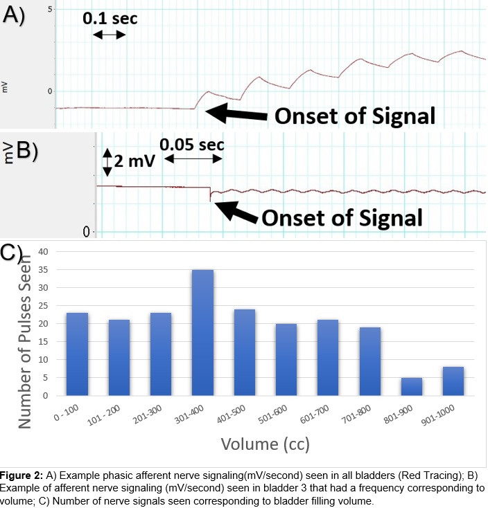
Results: Three porcine bladders were used for this study. In all animals a phasic afferent signal was identified (Figure 2A). Bursts of activity were noted at an average volume of 351 cc. The signal occurred over a period averaging 4 seconds with a frequency of 8 hz and amplitude of 0.65mV. An additional signal (Figure 2B) was observed (n=1) where signal activity corresponded to filling volume (Figure 2C). Nerves emanating from the posterior aspect of the bladder (n=1) were isolated and confirmed using H&E immunohistochemistry.
Conclusions: This experiment demonstrated that a perfused porcine bladder model can be used to identify afferent nerve signaling in response to filling. This is the first study of its type to use perfused porcine bladders. Application of this model to ongoing studies may be useful as an objective method to study bladder afferent function.
Source of Funding: NIH Grant R01DK101719


.jpg)
.jpg)