Back
Poster, Podium & Video Sessions
Moderated Poster
MP17: Pediatric Urology: Upper & Lower Urinary Tract
MP17-06: Evaluation of renal measurements of isolated high-grade congenital hydronephrosis (HGH) in both supine and prone positions
Friday, May 13, 2022
4:30 PM – 5:45 PM
Location: Room 225
Amr Hodhod*, Carolina Fermin-Risso, Mutaz Farhad, Anthony Cook, Jarrah Aburezeq, Steven Lu, Bryce Weber, Calgary, Canada

Amr Salah Hodhod, MSC
Alberta Children's Hospital
Poster Presenter(s)
Introduction: Some studies have questioned the optimal positioning for the assessment of urinary tract dilatation. Prone position offers better visualization of the kidneys in comparison to the supine position in which other abdominal organs can interfere with the ultrasound scan. In this study, we compared the ultrasound renal measurements in both prone and supine positions.
Methods: We conducted a retrospective review of patients presented with congenital isolated HGH, in the first year of life, from 2017-2019. A control group of patients with normal contralateral kidneys was included. The renal length, anteroposterior diameter of the renal pelvis (APD), parenchymal thickness (PT) and pyramidal thickness (PyT) were measured by a single investigator. APD was measured at the renal contour in the mid-renal transverse plane. Both PT and PyT were measured in the mid-zone of the renal sagittal plane. All measurements were obtained in both supine and prone positions.
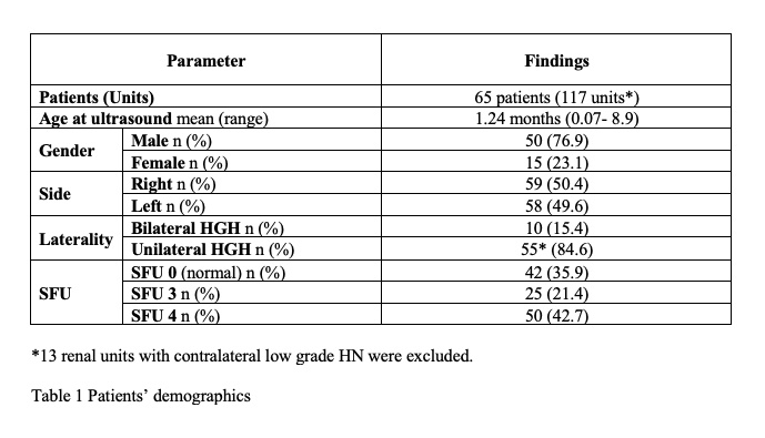
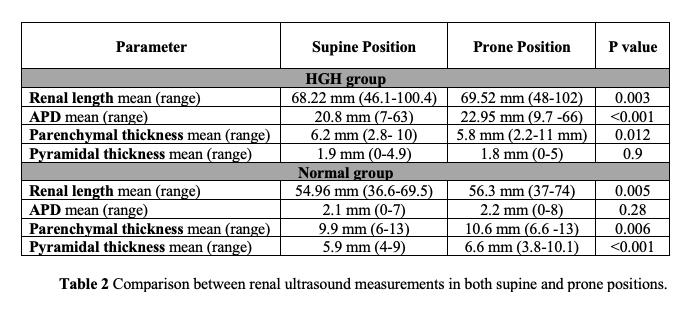
Results: Of 81 patients presented with congenital HGH, 65 patients (117 renal units) were included in our study. Contralateral normal forty-two renal units were included. Patients’ demographics are demonstrated in Table 1. Renal measurements in both positions are presented in Table 2. Prone measurements of renal length, in HGH and normal groups, were significantly higher (0.003, 0.005 respectively). In HGH group, although APD measurements in prone position exceeded those in the supine position (p < 0.001), PT measurements were significantly greater in the supine position (p=0.012). In the normal group, both PT and PyT measurements were lower in a supine position when compared to those in the prone position (p=0.006, <0.001 respectively).
Conclusions: There is a significant difference between renal measurements in both positions. However, the clinical impact of this difference should be further evaluated. Due to the prominence of hydronephrosis in the prone position, the PT and PyT measurements were less than the supine ones.
Source of Funding: None


.jpg)
.jpg)