Back
Poster, Podium & Video Sessions
Moderated Poster
MP18: Urodynamics/Lower Urinary Tract Dysfunction/Female Pelvic Medicine: Non-neurogenic Voiding Dysfunction I
MP18-18: Statistical Shape Modeling of Video Urodynamics: From Form to Function
Friday, May 13, 2022
4:30 PM – 5:45 PM
Location: Room 222
Kelsey Gallo*, Lindsey Burnett, San Diego, CA, Megan Routzong, La Jolla, CA, Marianna Alperin, Yahir Santiago-Lastra, San Diego, CA
.jpeg.jpg)
Kelsey Gallo, MD
UC San Diego Health
Poster Presenter(s)
Introduction: Video urodynamic studies (VUDS) assess LUT structure and function. Statistical shape modeling (SSM) defines principal components (PCs) that describe variations in shapes. Use of SSM to assess the bladder via imaging may detect previously undetectable alterations in bladder shape associated with dysfunction.
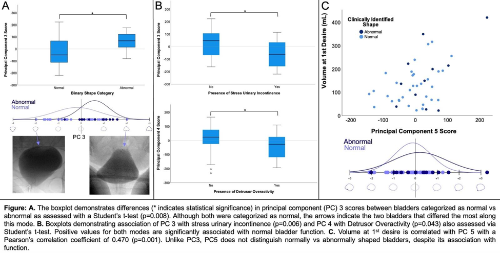
Methods: This IRB-approved retrospective study includes consecutive female participants with diagnoses of LUTS undergoing VUDS as part of their clinical care by a single provider. Fluoroscopic images of bladders at ~300ml were dichotomously categorized as normal or abnormal, defined as the presence of one or more bladder wall abnormalities (e.g., diverticuli, trabeculations). Bladders were digitally traced and converted to shapes defined by 400 landmarks. SSM was performed on the images by a researcher, who was blinded to clinical assessment of bladder shape. SSM is a principal component analysis (PCA) on aligned shape landmarks. SSM determined modes of variation in bladder shape. The main output of this is PCs defined by scores that are normally distributed about 0 and describe patterns of shape variation. Binary and continuous variables were assessed with appropriate statistical methods as detailed in figure.
Results: Bladder imaging was characterized as normal in 38/51 (74.5%) of participants. Despite this, all 51 had bladder functional abnormalities. SSM identified 9 PCs that describe variation in bladder shape. PCs were significantly associated with clinical phenotypes. PC3 (12% total shape variation) scores significantly differed across binary shape categories (Fig A) and between those with and without SUI (Fig B). PC4 (8% total shape variation) scores significantly differed in the presence/absence of DO (Fig B). PC 3 differed significantly for participants with normal vs abnormal bladder shapes. No other modes correlated to clinical assessment but many were associated with measures of bladder function. For example, PC 5 (4.5% total shape variation) correlated with volume at first desire.
Conclusions: SSM is a high resolution and robust technique that can be used to quantify subtle variation in bladder shape that are not identified clinically. These clinically undetectable alterations in bladder shape, quantified by PCs, may correspond with variation in bladder
Source of Funding: None


.jpg)
.jpg)