Back
Poster, Podium & Video Sessions
Best Poster Award
MP33: Uroradiology I
MP33-04: Contrast-Enhanced Ultrasonography for the Evaluation of Complex Renal Cysts
Saturday, May 14, 2022
4:30 PM – 5:45 PM
Location: Room 228
Cassra Clark*, Corinne Wessner, Shuo Wang, Andrew Denisenko, Andrew Shumaker, Joonyau Leong, Andrea Quinn, Erica Mann, Lydia Glick, Timothy Han, Kibo Nam, Katherine Smentkowski, John Eisenbrey, Leonard Gomella, Edouard Trabulsi, Costas Lallas, Mark Mann, James Mark, Flemming Forsberg, Andrej Lyshchik, Ethan Halpern, Thenappan Chandrasekar, Philadelphia, PA

Cassra B. Clark, MD
Urology Research Fellow
Sidney Kimmel Medical College at Thomas Jefferson University
Poster Presenter(s)
Introduction: Complex renal cysts are evaluated using the Bosniak classification system. However, they remain difficult to manage because inter-reader variability and subjective interpretation of the Bosniak system. This pilot study intends to evaluate the use of contrast-enhanced ultrasonography (CEUS) to differentiate the solid and cystic components of complex renal cysts and compare these results to the final surgical pathology report.
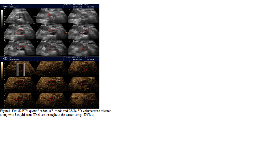
Methods: 23 patients undergoing surgery for Bosniak 2F-4 lesions participated in this IRB-approved pilot study. Patients were scanned prior to surgery with both 2D and 3D ultrasound exams. Each injection consisted of a 2.0 mL bolus injection of Lumason contrast (Bracco Imaging, Monroe Township, NJ) followed by a 10 mL saline flush. A custom MATLAB program was used for selection of regions of interest (ROIs) (Figure 1). Fractional tumor vascularity (FTV in %) was then calculated and compared to estimated solid component on the final pathology report.
Fractional Tumor Vascularity = 1 – (Total Non-enhancing area/Total lesion area)
The primary endpoint of this study was the correlation of 2D/3D-derived fractional vascularity with pathological estimation and tumor staging on explant.
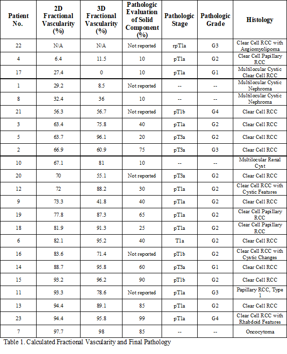
Results: No adverse events were observed during any contrast agent administration. Data was analyzed on 22 of 23 patients (Patient 22 excluded due to poor image quality). Mean age was 60.9 ± 15.0 years (range 30 to 85) and mean preoperative lesion size was 4.0 ± 1.6 cm (range 1.2 to 8.3 cm). Nine patients had radical nephrectomies and 14 had a partial nephrectomy. On final pathology, 4 lesions were benign and 19 were malignant. The results of 2D and 3D analysis as well as final pathology are included in Table 1. In general, malignant lesions and more aggressive histology had higher FTV.
Conclusions: Quantitative 2D and 3D CEUS can evaluate FTV of potentially malignant complex cystic renal masses, which may be able to augment current radiographic classification systems.
Source of Funding: N/A


.jpg)
.jpg)