Back
Poster, Podium & Video Sessions
Moderated Poster
MP33: Uroradiology I
MP33-11: Development of a Urinary Tract Atlas Using Convolutional Neural Networks
Saturday, May 14, 2022
4:30 PM – 5:45 PM
Location: Room 228
Katherine Fischer*, Yuemeng Li, Benjamin Schurhamer, Justin Ziemba, Yong Fan, Gregory Tasian, Philadelphia, PA

Katherine M. Fischer, MD
Children's Hospital of Philadelphia
Poster Presenter(s)
Introduction: The management of urolithiasis relies heavily on the features of the stone and surrounding urinary anatomy on CT. Current practice patterns utilize human interpretation to identify and assess genitourinary anatomical features, which is laborious and poorly reproducible. Automated analysis of CT images to perform these tasks can potentially increase accuracy, fidelity, and speed. We developed a convolutional neural network model to automatically segment the kidneys, ureters, and bladder on CT, thereby creating an atlas of the urinary tract.
Methods: A total of 29 CT urograms (CTU) from adult patients undergoing imaging for nephrolithiasis were utilized. The scans were manually segmented to outline each kidney, ureter, and the bladder. A standard U-Net model was utilized containing 5 encoder blocks, a bottleneck block, and 5 decoder blocks. Each block contained two convolution layers with a kernel size of 3. The maxpooling function was applied at the end of each encoder block and the up-sampling function was applied at the end of each decoder block. All the CT scans were resampled to the spatial dimension of (2.5, 1.62, 1.62) with respect to axial, sagittal, and coronal views. We clipped the CT scan intensity to the range [-200, 450]. The network was trained based on the input patches size of (96, 160, 160). We randomly split the data into a training set of 25 CTUs and testing set of 4 CTUs.
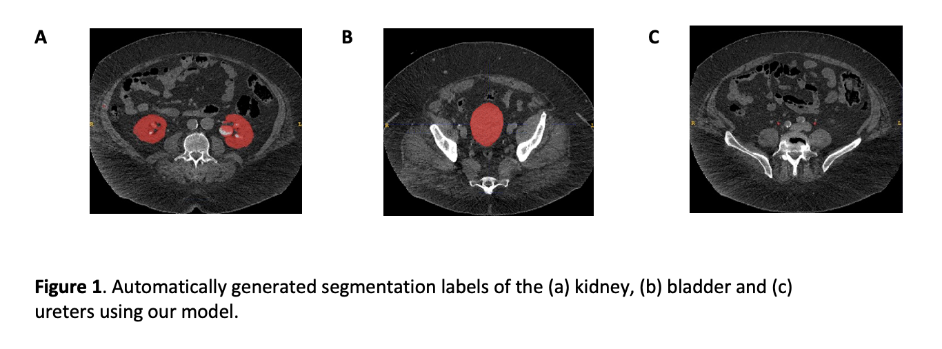
Results: A convolutional neural network model was developed that automatically segmented the kidneys, ureters, and bladder (Figure 1). Dice scores were 0.644±0.388 for bladder segmentation, 0.879±0.058 for kidney segmentation, and 0.610±0.101 for ureter segmentation.
Conclusions: We created a machine learning model that automatically segmented the urinary tract with moderate to high precision. Our next step is to improve model performance by increasing the number of images in the training set. Ultimately, this atlas will be utilized to allow prediction of the probability of ureteral stone passage in children and adults. Automation of CT imaging analysis has the potential to usher in personalized medicine for patients with kidney stone disease where passage of ureteral stones and other outcomes could be predicted at the time of diagnosis.
Source of Funding: This project was supported by NIH grant P20DK127488.
This work was supported in part by the 2021-2022 Urology Care Foundation Research Scholar Award Program (KF).


.jpg)
.jpg)