Back
Poster, Podium & Video Sessions
Moderated Poster
MP47: Kidney Cancer: Epidemiology & Evaluation/Staging/Surveillance III
MP47-05: Stimulated Raman Histology Allows for Real Time Determination of the Adequacy of Renal Mass Biopsy and Identify Malignant Subtypes of Renal Cell Carcinoma
Sunday, May 15, 2022
2:45 PM – 4:00 PM
Location: Room 228
Miles P. Mannas*, Fang-Ming Deng, Derek Jones, William Huang, Daniel Orringer, Samir S Taneja, New York, NY

Miles Mannas, MD, FRCSC
NYU Langone Health
Poster Presenter(s)
Introduction: Renal biopsy requires adequate tissue sampling to aid in the investigation of renal masses. The contemporary rate of non-diagnostic renal biopsy ranges from 15-18%, though may be as high as 42% in challenging cases. Simulated Raman Histology (SRH) is a novel microscopic technique which has created the possibility for rapid, label-free, high-resolution images of unprocessed tissue which may be viewed on standard radiology viewing platforms. The application of SRH to renal biopsy may provide the benefits of pathologic hematoxylin and eosin (H&E) evaluation during the procedure, thereby reducing non-diagnostic results. We conducted a pilot feasibility study, to assess if all RCC subtypes may be imaged and to see if high-quality H&E could subsequently be generated.
Methods: An 18-gauge core needle biopsy was taken from a series of 9 ex vivo radical or partial nephrectomy specimens. Histologic images of the fresh, unstained biopsy samples were obtained using a SRH microscope using two Raman shifts: 2845cm-1 and 2930cm-1. The cores were then processed as per normal pathologic protocols. The SRH images and H&E were then viewed by a genitourinary pathologist.
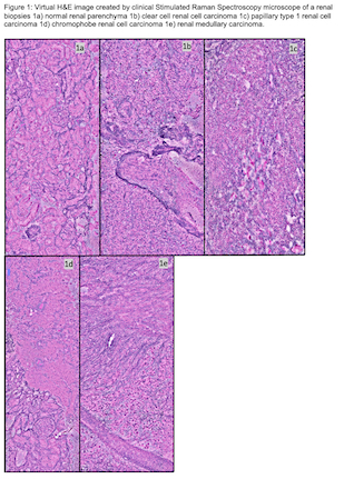
Results: The SRH microscope took 8-11 minutes to produce high-quality images of the renal biopsies. All renal cell carcinoma subtypes were captured, and the images are shown in figure 1. SRH imaged biopsies were interpreted by a genitourinary pathologist and areas of tumor were easily differentiated from normal renal parenchyma. Figure 1a shows SRH of normal renal parenchyma, while figure 1b-e shows SRH for clear cell-, papillary-, chromophobe renal cell carcinoma and renal medullary carcinoma. High quality H&E was produced from each of the renal biopsies after SRH was completed.
Conclusions: SRH produces high quality images of all renal cell subtypes that can be rapidly produced and easily interpreted to determine renal mass biopsy adequacy and may allow improved renal cell carcinoma subtype identification. Renal biopsies remained available to produce high quality H&E for confirmation of diagnosis. Procedural application has promise to decrease the rate of renal mass non-diagnostic renal mass biopsies and application of convolutional neural network methodology may improve diagnostic capability for biopsy of renal masses.
Source of Funding: NIH Grant: UL1TR001445


.jpg)
.jpg)