Back
Poster, Podium & Video Sessions
Moderated Poster
MP51: Prostate Cancer: Staging
MP51-06: PIRADS score combined with positive core number on MRI-ultrasound fusion targeted prostate biopsy can predict the absence of pathological extraprostatic extension
Sunday, May 15, 2022
4:30 PM – 5:45 PM
Location: Room 222
Masaki Kobayashi*, Yoh Matsuoka, Sho Uehara, Hiroshi Tanaka, Yuki Nakamura, Yusuke Uchida, Shohei Fukuda, Hajime Tanaka, Soichiro Yoshida, Minato Yokoyama, Yasuhisa Fujii, Tokyo, Japan
Poster Presenter(s)
Introduction: The prediction of pathological extraprostatic extension (EPE) is crucial in selecting treatment, especially for neurovascular bundle (NVB) preservation in radical prostatectomy (RP). EPE assessment is highly dependent on morphological MRI findings. However, whether or not prostate cancer (PC) with capsular abutment on MRI is suitable for NVB preservation is unclear. Today, the role of a targeted biopsy (TB) in predicting EPE is also unclear. We thus evaluated the utility of MRI and TB findings for prediction of EPE and clarified when NVB preservation is possible in PC with capsular abutment.
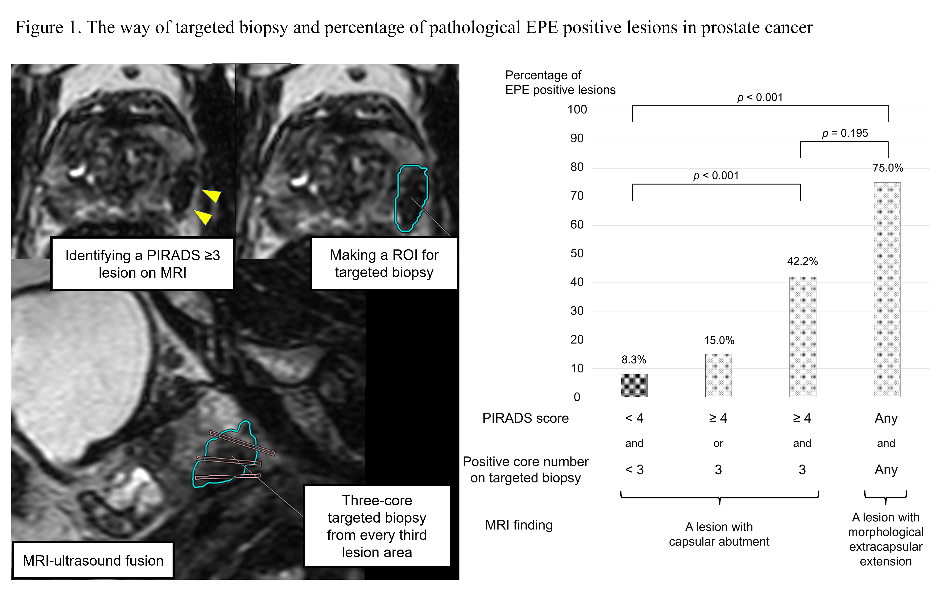
Methods: We enrolled 120 consecutive patients undergoing an MRI-ultrasound fusion TB and systematic biopsy followed by robot-assisted RP (IRB #M2000-2130-01). All MR images were interpreted by an experienced radiologist. We performed 3-core TBs of each PIRADS =3 lesion by obtaining each core from every third lesion area. We checked each lesion for EPE by referencing RP specimens and explored EPE-predictive MRI and TB variables. ROC curves were used to decide cutoff value.
Results: We identified 206 PIRADS =3 lesions, including 174 with capsular abutment, 12 with morphological extracapsular extension, and 20 without capsular contact. Among 174 lesions with capsular abutment, 64, 74, and 36 had PIRADS 3, 4, and 5, respectively. On TBs, cancer was detected in 120 lesions (69%). In RP specimens, EPE was found in 35 of 174 lesions (20%) with capsular abutment and 9 of 12 (75%) with morphological extracapsular extension. A univariate analysis of 174 lesions with capsular abutment showed that EPE was associated with PIRADS =4 (p=0.01), Gleason score =3+4 (p=0.01), 3 positive cores (p < 0.01), and maximum cancer length =5 mm (p < 0.01) on a TB. In the multivariate analysis, PIRADS =4 (odds ratio [OR] 3.0, p=0.03) and 3 positive cores (OR 3.3, p<0.01) were independent predictors of EPE. EPE was found in 8% of PIRADS <4 lesions with <3 positive cores and 42% of PIRADS =4 lesions with 3 positive cores (p < 0.01).
Conclusions: The combination of the PIRADS score and positive core number on a TB were useful for predicting pathological EPE in PC with capsular abutment. NVB can be preserved on the site with PIRADS <4 lesions in which limited number of positive cores are detected on a TB.
Source of Funding: No funding.


.jpg)
.jpg)
