Back
Poster, Podium & Video Sessions
Moderated Poster
MP53: Prostate Cancer: Detection & Screening V
MP53-06: MRI and MRI-targeted biopsy can detect cribriform cancer of the prostate
Monday, May 16, 2022
7:00 AM – 8:15 AM
Location: Room 225
Sho Uehara*, Yoh Matsuoka, Kurara Yamamoto, Yuki Nakamura, Yusuke Uchida, Shohei Fukuda, Hajime Tanaka, Soichiro Yoshida, Minato Yokoyama, Kenichi Ohashi, Yasuhisa Fujii, Tokyo, Japan
- SU
Sho Uehara, MD
Tokyo Medical and Dental University Graduate School
Poster Presenter(s)
Introduction: Cribriform cancer (CC) of the prostate has been recognized as an aggressive entity. To date, whether cribriform component is less visible on multiparametric MRI (mpMRI) than other Gleason pattern 4 cancer or not is controversial. In addition, diagnostic performance of MRI-ultrasound fusion targeted biopsy (TB) for CC detection has not been well investigated. The aim of our study is to examine the visibility of CC on mpMRI and CC detection by TB, in reference to systematic biopsy (SB) and robot-assisted radical prostatectomy (RARP) findings.
Methods: Consecutive 103 prostate cancer patients were investigated who underwent RARP without neoadjuvant treatment. All patients had undergone TB concurrently with 12-core SB at diagnosis. PIRADS v2.1 scores were prospectively assigned prior to biopsy. The detection rate of CC and significant cancer (SC) on TB, SB, and RARP was examined. SC was defined as cancer with grade group =2.
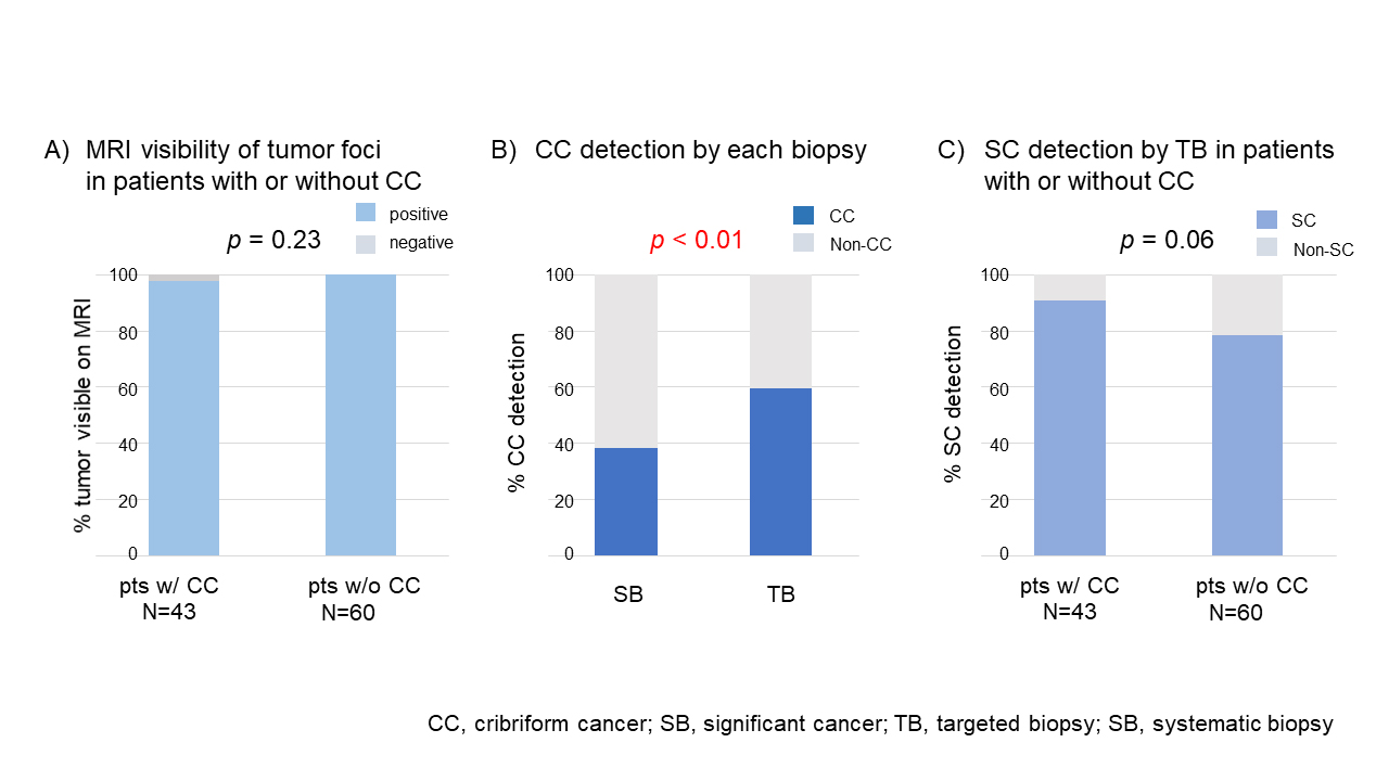
Results: Median age, PSA, and prostate volume were 68 years, 9.0 ng/mL, and 29.7 mL, respectively. Median volume of index lesions was 0.72 mL and their PIRADS scores were 3/4/5 in 18/40/45 patients, respectively. In RARP specimens of 103 patients, CC and SC were found in 43 (42%) and 98 (95%) patients, respectively. On mpMRI, CC foci in RARP specimens were visible in 42 patients (98%) and scored as PIRADS 3/4/5 in 5/16/21patients, respectively. In one patient, CC was not identified as abnormal lesion on mpMRI. CC were identified by SB and TB in 15(35%), 24(56%) patients, respectively (SB vs TB, p = 0.01), and SC were detected by SB and TB in 33 (77%), 39 (91%) patients, respectively (SB vs TB, p = 0.01). In the remaining 60 patients without CC on RARP specimens, PIRADS score 3/4/5 were assigned in 13/24/23 patients. SC were detected by SB and TB in 33 (55%) and 47 (78%) patients, respectively (SB vs TB, p <0.01). MRI visibilities of prostate cancer were 98% in patients with CC and 100% in patients without CC. TB missed SC in 4 of 43 patients with CC and in 13 of 60 patients without CC (p=0.10).
Conclusions: Majority (98%) of CC foci were visible on mpMRI, and CC were identified by TB in more than half (56%) of the patients with CC. SC detection by TB did not differ between patients with CC and those without CC.
Source of Funding: None


.jpg)
.jpg)