Back
Poster, Podium & Video Sessions
Benign Prostatic Hyperplasia: Basic Research & Pathophysiology
PD16-02: Ultrastructural Findings of COVID-19 Infection in the Prostate
Friday, May 13, 2022
3:40 PM – 3:50 PM
Location: Room 244
Rohit Reddy*, Eliyahu Kresch, Natoli Farber, Parris Diaz, Ranjith Ramasamy, Miami, FL

Rohit Reddy, MD, BA
Temple University Hospital
Podium Presenter(s)
Introduction: SARS-CoV-2 utilizes two integral membrane proteins ACE2 and TMPRSS2 for viral replication. It has been established TMPRS22 specifically is found in high concentrations throughout the prostate found to be linked to prostatic disease progression. This project examined the histopathological, ultrastructural, and immunofluorescent elements of prostatic tissue from men infected by SARS-CoV-2.
Methods: We evaluated prostate tissue in men with worsening lower urinary tract symptoms who underwent HoLEP procedure after SARS-CoV-2 infection. Biopsied tissue was visualized by transmission electron microscopy (TEM), immunofluorescence, and viral presence was confirmed by quantitative polymerase chain reaction (RT-PCR).
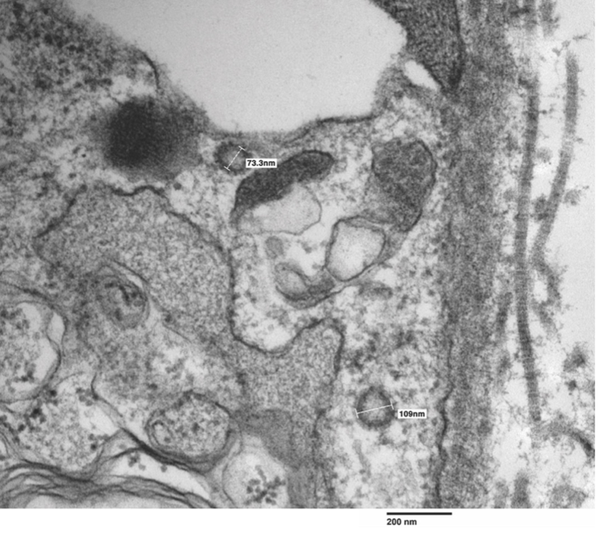
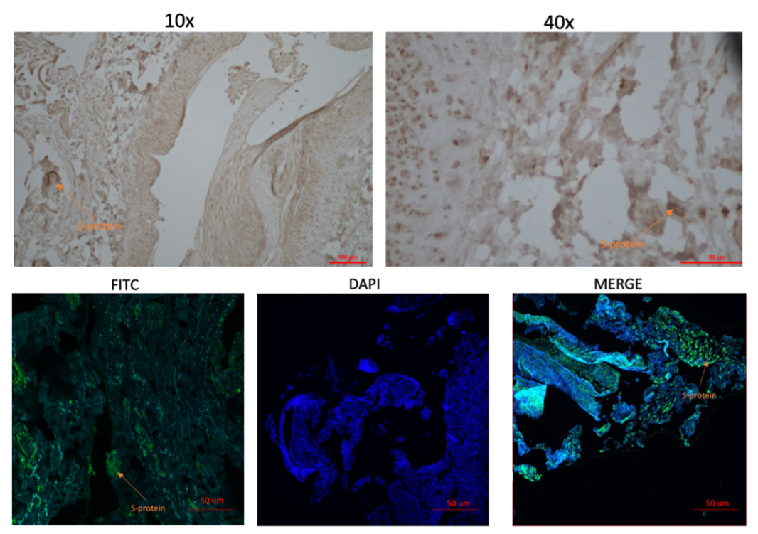
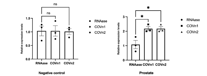
Results: Multiple coronavirus-like spiked viral particles ranging from 73.3mm to 109mm were visualized by TEM (Figure). Histochemical and immunofluorescence concurrently showed presence of distinct hyalinization, fibrosis, and presence of spike protein (Figure 2). RT-PCR confirmed the identity of the viral bodies as SARS-CoV-2 (Figure 3).
Conclusions: This study provides evidence that SARS-CoV-2 not only enters prostatic tissue but may persist beyond initial infection period. In addition to establishing the persistence of SARS-CoV-2 particles in prostatic tissue, this report suggests the importance of discerning the relationships between COVID-19, lower urinary tract symptom severity, and prostatic hyperplasia.
Source of Funding: This work was supported by National Institutes of Health Grant R01 DK130991 and Clinician Scientist Development Grant from the American Cancer Society to RR.


.jpg)
.jpg)