Back
Poster, Podium & Video Sessions
Podium
PD27: Prostate Cancer: Localized: Surgical Therapy II
PD27-01: Predicting Extra-prostatic Extension (EPE) for surgical guidance in prostate cancer: A comparison of biopsy pathology, multiparametric MRI, and PSMA-PET
Saturday, May 14, 2022
1:00 PM – 1:10 PM
Location: Room 255
Clinton Bahler*, Mark Green, Mark Tann, Katrina Collins, Jordan Swensson, Eric Brocken, Liang Cheng, Carla Mathias, Indianapolis, IN, David Alexoff, Hank Kung, Philadelphia, PA, Gary Hutchins, Michael Koch, Indianapolis, IN

Clinton D. Bahler, MD
Indiana University
Podium Presenter(s)
Introduction: Prostatectomy related incontinence and impotency result from injury to nerves and muscle tissue. Preserving nerves and muscle adjacent to the prostate risks positive margins; therefore, preoperative imaging is needed with high sensitivity for extra-prostatic extension.
Methods: Four clinical trials for PSMA-PET targeted imaging prior to prostatectomy were retrospectively evaluated for prediction of extra-prostatic extension as documented on post-surgical whole-mount pathologic analysis. Two different PSMA-PET tracers were included: PSMA-11 and P16-093. A blinded review of PET and MRI scans was performed to predict the risk of extra-prostatic extension (EPE). Stata 13.1 was used. Pearson’s Chi2 and McNemar’s Chi2 were used for accuracy statistics.
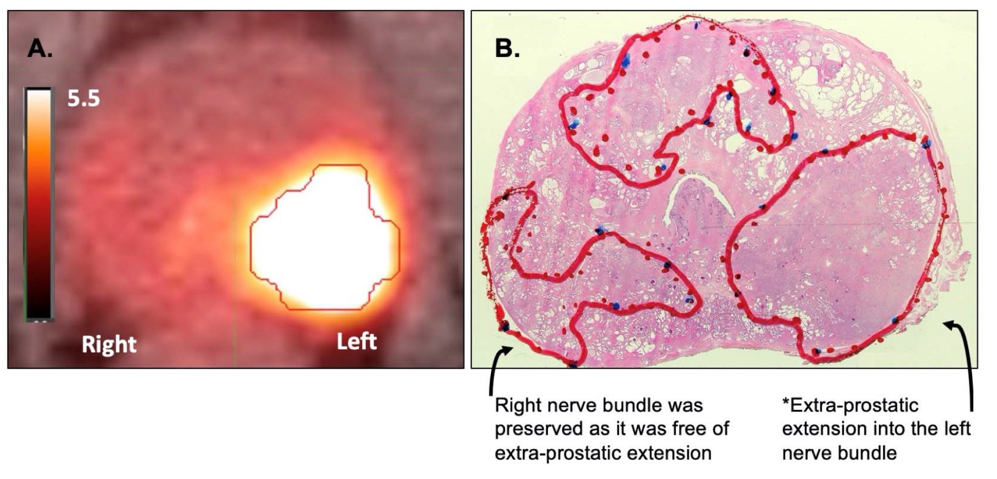
Results: Pre-operative PSMA-PET imaging was performed in 71 patients with either 68Ga-P16-093 (n=25) or 68Ga-PSMA-11 (n=46). Age (62 vs 61 years), PSA (7.3 vs 7.8), and biopsy pathology (3+3: 0 vs 7%, 3+4: 28 vs 39%, 4+3: 24 vs 20%, =4+5: 48 vs 35%) were similar between the groups (p>0.30). There were 24 (34%) with pT3a (EPE) and 16 (23%) with pT3b on final pathology. Overall positive margin rate was 17 (24%). Index lesion SUVmax was similar between P16-093 and PSMA-11 (10.3 vs 11.7). EPE Sensitivity (87 vs 92%), Specificity (77 vs 76%), and ROC area (82 vs 84%) were similar between P16-093 and PSMA-11, respectively (p=0.87). MRI (available in 45) found high specificity (83%) but low sensitivity (60%) and the overall ROC area was lower when compared to pooled PSMA-PET (0.72 vs 0.83, p=0.01). For pooled PSMA-PET imaging, a treatment change from “non-nerve sparing” to “nerve sparing” was recommended in 21/71 (30%) of patients. A total of 14 men had nerve-sparing (treatment change) based on PSMA-PET imaging with one (5%) having a positive margin. Figure 1 shows a case where left-sided EPE as accurately predicted by 68Ga-P16-093 scan.
Conclusions: PSMA-PET imaging can improve surgical guidance in men with =4+3 prostate cancer resulting in preservation of nerve-bundles. 68Ga-P16-093 PET and 68Ga-PSMA-11 PET had similar accuracy for predicting extra-prostatic extension.
Source of Funding: Al Christy Prostate Cancer Fund, R44 CA233140, NIH Small Business Innovation Research grant (SBIR), CTSI NIH/NCRR Grant Number UL1TR001108, ACS-IRG Grant Mechanism (16-192-31)


.jpg)
.jpg)