Back
Poster, Podium & Video Sessions
Podium
PD28: Sexual Function/Dysfunction: Surgical Therapy I
PD28-01: How accurate are we when placing submuscular reservoirs during multi-component penile implant surgery
Saturday, May 14, 2022
1:00 PM – 1:10 PM
Location: Room 245
Bruce Kava*, Amanda Levine, Thomas Masterson, Nicholas Hauser, Ranjith Ramasamy, MIami, FL

Bruce Richard Kava, MD
University of Miami Miller School of Medicine
Podium Presenter(s)
Introduction: Techniques that provide for sub-muscular placement of prosthetic reservoirs fulfil a critical need for those patients who have undergone prior pelvic surgery and who are desirous of a multicomponent inflatable penile prosthesis. Contemporary approaches rely on passage of the reservoir through the inguinal canal or directly through the anterior rectus fascia. Studies using cadavers have shown that approximately 65% of these reservoirs are placed unknowingly within the peritoneum, retroperitoneum, or lateral abdominal wall. Our objective was to use post- implant cross sectional imaging to ascertain how often the reservoir was located within the correct sub-muscular location.
Methods: The cohort of patients was obtained from an IRB- approved database of consecutive patients undergoing penile prosthesis surgery. Patients undergoing sub-muscular reservoir placement who underwent cross- sectional imaging (MRI or CT scan) represented our study cohort. The reason for undergoing imaging was abstracted and the scans were reviewed to determine the post-implant location of the reservoir.
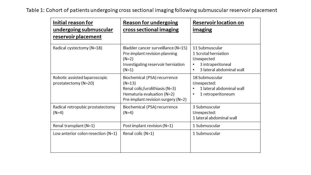
Results: Cross- sectional imaging was performed in 43 patients. Table 1 summarizes the reasons for pursuing abdominal imaging, and the location of the reservoir. There were 9 (21%) reservoirs in unexpected locations, including 5 (12%) devices within the lateral abdominal musculature, 1 (2%) device anterior to the psoas fascia, and 3 (7%) within the peritoneal cavity. None of these patients was symptomatic.
Conclusions: Despite careful and deliberate sub-muscular reservoir placement, 20% of these reservoirs will be placed unknowingly within the lateral abdominal wall, the retroperitoneum, and the peritoneal cavity. While the lateral abdominal wall poses little threat to the patient, intraperitoneal and retroperitoneal reservoirs may subsequently lead to the development of bowel obstruction or fistulas. Therefore preoperative counseling should include a discussion of these potential risks, particularly in post- cystectomy patients. Additionally, preoperative imaging may be a consideration for those patients undergoing revision surgery in whom the reservoir will either be drained and retained or left in situ. This would enable intraperitoneal reservoirs to be identified and be considered for removal.
Source of Funding: None


.jpg)
.jpg)