Back
Poster, Podium & Video Sessions
Podium
PD37: Surgical Technology & Simulation: Instrumentation & Technology II
PD37-06: Comparison of Computational simulation and Hydrogel kidney phantoms for in vivo assessment of Intrarenal pressure (IRP) dynamics during ureteroscopy under various experimental conditions.
Sunday, May 15, 2022
7:50 AM – 8:00 AM
Location: Room 245
Ahmed Ghazi*, Rachel Melnyk, Andrew Cook, Nitin Sharma, Hana kalco, Tran Phuc, David Foster, Rochester, NY

Ahmed E. Ghazi, MD, FEBU, MBBS
University of Rochester
Podium Presenter(s)
Introduction: Several in vivo studies have analyzed methods of mitigation of high Intrarenal pressure (IRP) to reduce the risk of pyelovenous backflow. These studies have neglected the effect of stones on the flow dynamics to different areas within the pelvicaleceal system (PCS). Herein, we evaluated two in vivo simulation methods [computational fluid dynamics (CFD) and anatomical patient specific phantoms] with and without the inclusion of stones.
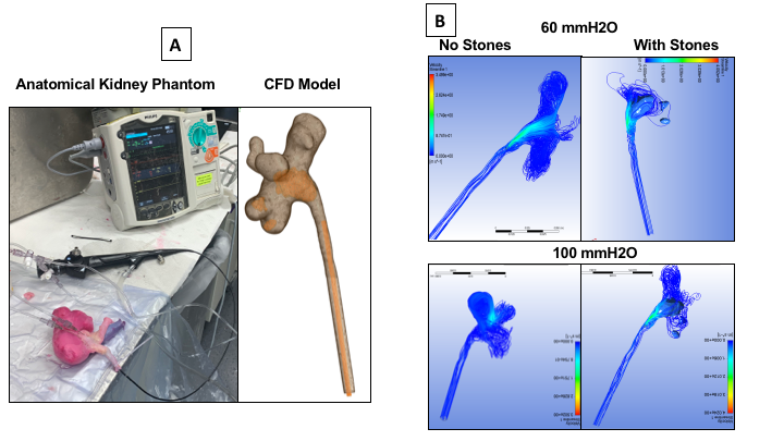
Methods: A computer design was created from a patients CT urogram with a 1.8 cm pelvic 7 and 5mm lower pole stones. A kidney phantom and CFD model were fabricated (Fig 1A). CFD iteratively solves equations governing fluid dynamics resulting in millions of data points used to simulate the physiological flow and measure pressures of body fluids. Hydrogel phantoms were constructed of patient’s PCS and kidney, with and without the stones using our published techniques for replicating realistic mechanical properties where pressure data was collected from each PCS location using percutaneous sensors (Fig1). Two independent teams completed simulations where a uretereoscope (Olympus URF-P6) was physically positioned at the tip of the pelvic stone. IPP was collected from each models at locations within the PCS, at low (60 cmH20) and high (100 cmH20) inflow rates both with and without the stones.
Results: In the CFD model, an mean difference of 6.2 and 7.3 mmH2O was seen at 60 and 100mmH2O inflow when including the stones with the largest change occurring at the lower pole (Fig 1B). For the hydrogel model, mean difference of 16 and 20 mmH2O was seen at 60 and 100mmH2O inflow when including stones with the largest change also occurring at the lower pole . Directly comparing the models will require further alignment between models to settle differences in scope placement and orientation.
Conclusions: This is the first study to demonstrate that stones significantly change fluid dynamics during ureteroscopy using patient specific CFD and validated hydrogel models, a departure from previous studies. Future investigation will investigate the factors contributing to the differences between the models and explore how variation in patient anatomy effects fluid flow and pressures.
Source of Funding: NA


.jpg)
.jpg)